Endocrine System Physiology and Hormonal Regulation
An in-depth explanation of the endocrine system, covering hormone release mechanisms, feedback loops (negative and positive), the hypothalamic-pituitary axis, cellular mechanisms of hormone action, and common endocrine disorders.
The endocrine system is a sophisticated communication network that utilizes chemical messengers, known as hormones, to maintain homeostasis—the stable internal environment required for survival. Unlike the nervous system, which uses electrical impulses for rapid, localized responses, the endocrine system typically operates through the bloodstream, allowing for slower but more widespread and long-lasting effects on target tissues. To maintain this balance, the system relies on complex regulatory loops driven by specific stimuli and feedback mechanisms.
Mechanisms of Hormone Release
Hormones do not enter the bloodstream at random; their secretion is tightly controlled by three distinct types of stimuli:
Humoral Stimuli: This refers to the monitoring of blood levels of specific ions or nutrients. For example, when blood calcium levels drop, the parathyroid glands detect this change and release parathyroid hormone (PTH) to restore balance. Similarly, rising blood glucose levels after a meal act as a humoral stimulus for the pancreas to release insulin.
Hormonal Stimuli: This occurs when one hormone triggers the release of another. This is the foundation of the "axis" concept. For instance, the hypothalamus secretes Thyrotropin-Releasing Hormone (TRH), which travels to the anterior pituitary to stimulate the release of Thyroid-Stimulating Hormone (TSH).
Neural Stimuli: The nervous system can directly trigger endocrine responses. A classic example is the "fight-or-flight" response, where sympathetic neurons stimulate the adrenal medulla to immediately release epinephrine (adrenaline) into the blood.
The Architecture of Feedback Loops
Every homeostatic response follows a four-part functional loop: a stimulus (the environmental change), a sensor/receptor (the detection of that change), a control center (the processing unit, often the brain or a gland), and an effector (the organ that carries out the response).
Negative Feedback: The Stabilizer
Negative feedback is the most common regulatory mechanism in the body. It functions like a thermostat: once the desired physiological state is achieved, the system "shuts off" the initial stimulus to prevent overcorrection.
A primary example is blood glucose regulation:
High Glucose: The pancreas (sensor/control center) releases insulin (effector), which promotes glucose uptake by cells, lowering blood sugar.
Low Glucose: The pancreas releases glucagon (effector), which signals the liver to break down glycogen into glucose, raising blood sugar.
Another critical example is the Hypothalamic-Pituitary-Adrenal (HPA) axis, which manages the stress response:
The hypothalamus releases Corticotropin-Releasing Hormone (CRH).
CRH stimulates the anterior pituitary to release Adrenocorticotropic Hormone (ACTH).
ACTH stimulates the adrenal cortex to release cortisol.
The Shutoff: Once cortisol levels rise, the cortisol itself acts as a negative feedback signal, inhibiting the release of both CRH and ACTH to prevent excessive cortisol production.
Positive Feedback: The Amplifier
Positive feedback is rare because it moves the body away from homeostasis to achieve a specific, explosive climax. Instead of reversing a change, it amplifies it.
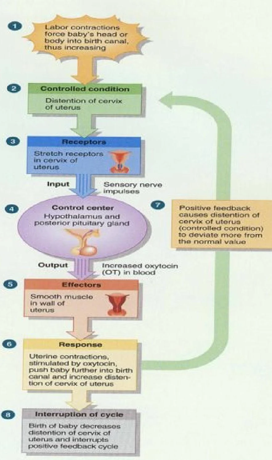
The most famous example is oxytocin during childbirth:
The baby's head pushes against the cervix (stimulus).
Nerve impulses signal the brain to release oxytocin (effector).
Oxytocin causes uterine contractions, which push the baby harder against the cervix.
This increased pressure triggers even more oxytocin, creating a loop that only breaks once the baby is delivered.
This diagram illustrates the positive feedback loop of labor contractions, involving the cervix, hypothalamus, pituitary gland, and oxytocin, leading to childbirth and the interruption of the cycle.
The Hypothalamic-Pituitary Axis
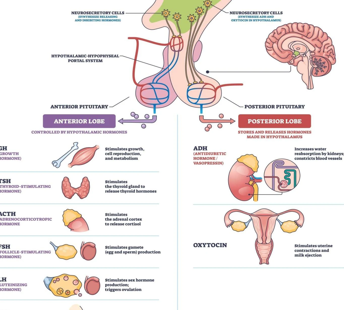
The hypothalamus acts as the "master regulator," serving as the bridge between the nervous and endocrine systems. It controls the pituitary gland, which is divided into two distinct functional lobes.
The Anterior Pituitary (Adenohypophysis)
The anterior pituitary is controlled by hypothalamic "releasing" or "inhibiting" hormones that travel through a specialized blood vessel network called the hypophyseal portal system. It secretes tropic hormones, which are hormones that target other endocrine glands:
TSH (Thyroid-Stimulating Hormone): Targets the thyroid to release T3 and T4, regulating metabolism.
ACTH (Adrenocorticotropic Hormone): Targets the adrenal cortex to release cortisol.
FSH & LH (Gonadotropins): Target the gonads (testes/ovaries) to regulate reproduction.
GH (Growth Hormone): Targets the liver to produce IGF-1 and acts on bones/muscles for growth.
PRL (Prolactin): Targets mammary glands to stimulate milk production.
This diagram illustrates the human endocrine system, focusing on the pituitary gland and its hormonal regulation of various bodily functions, including growth, metabolism, thyroid function, adrenal function, gamete production, and uterine contractions.
The Posterior Pituitary (Neurohypophysis)
Unlike the anterior lobe, the posterior pituitary does not synthesize its own hormones. Instead, it stores and releases hormones produced in the hypothalamus:
ADH (Antidiuretic Hormone / Vasopressin): Targets the kidneys to increase water reabsorption, helping to maintain blood pressure and fluid balance.
Oxytocin: Targets the uterus and mammary glands for contractions and milk ejection.
Diagram illustrating the relationship between hypothalamic releasing hormones and anterior pituitary hormones, detailing their targets and effects on various bodily systems.
Major Regulatory Axes
The integration of these glands forms specific pathways known as "axes":
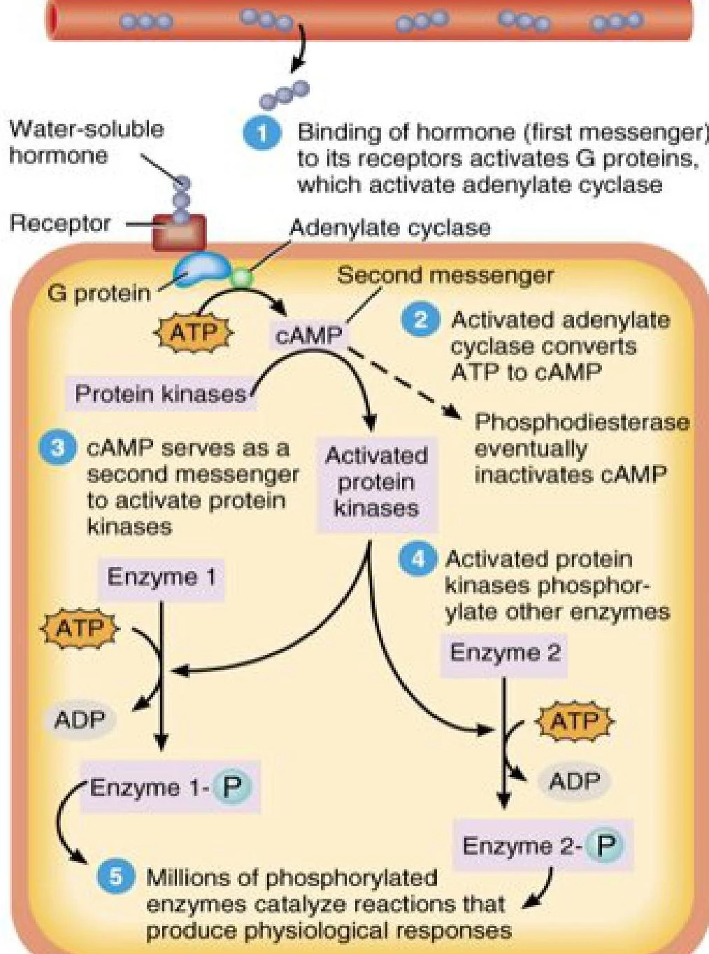
Hydrophilic hormones, such as peptide hormones (insulin, glucagon) and amino acid derivatives (epinephrine), cannot cross the fatty cell membrane.
Mechanism: The hormone (the "first messenger") binds to a receptor on the outside of the cell membrane. This binding activates a G-protein, which in turn activates an enzyme (like adenylate cyclase) to produce a second messenger (like cyclic AMP or cAMP) inside the cell.
Result: The second messenger triggers a rapid cascade of biochemical reactions, such as activating protein kinases to change cellular metabolism.
This diagram illustrates the mechanism of signal transduction initiated by a water-soluble hormone binding to its receptor, leading to a cascade of intracellular events involving G proteins, cAMP, and protein kinases, ultimately resulting in physiological responses.
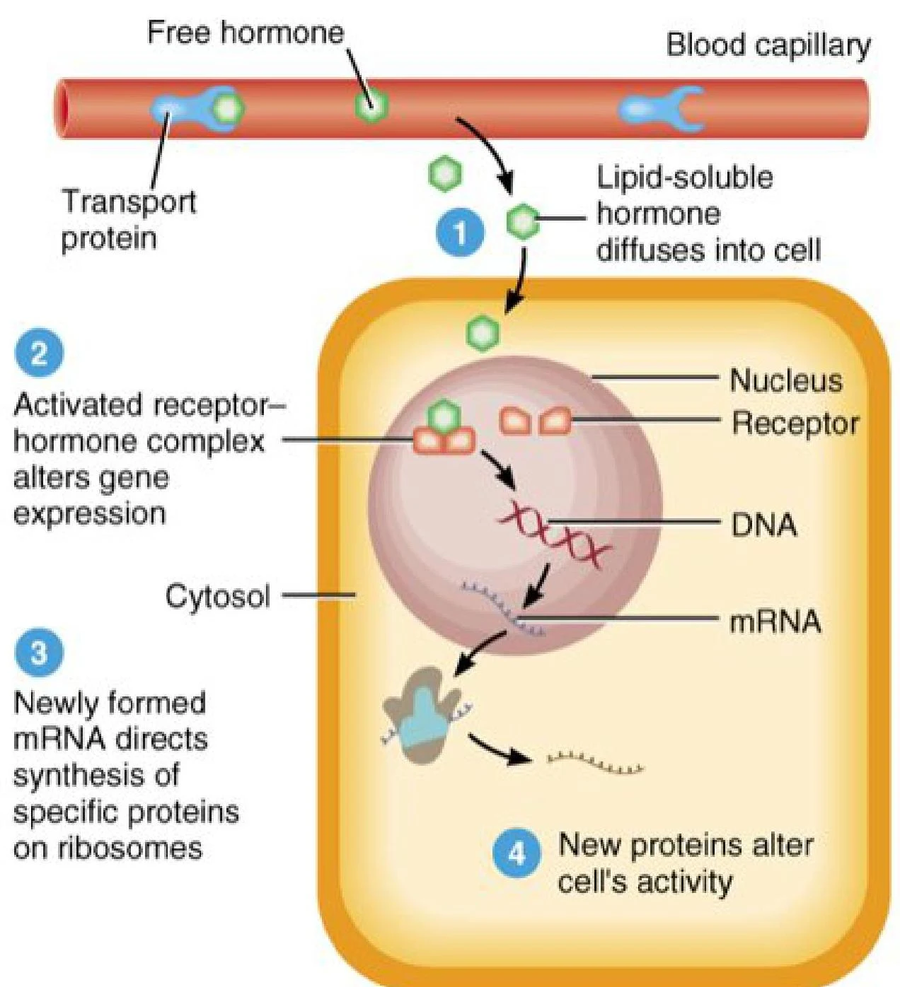
Lipophilic hormones, such as steroid hormones (cortisol, testosterone, estrogen) and thyroid hormones (T3/T4), can diffuse directly through the plasma membrane.
Mechanism: Once inside, they bind to receptors in the cytoplasm or the nucleus. The hormone-receptor complex then binds directly to the cell's DNA.
Result: This acts as a transcription factor, turning specific genes "on" or "off" to synthesize new proteins, leading to long-term changes in cell activity.
This diagram illustrates the mechanism of action for lipid-soluble hormones. A free hormone binds to a receptor inside the cell, forming a complex that alters gene expression. This leads to the synthesis of new proteins on ribosomes, which then modify the cell's activity.
Clinical Significance and Disorders
Endocrine disorders arise when the delicate balance of secretion or reception is disrupted. These are generally categorized by the nature of the secretion: hypersecretion (too much) or hyposecretion (too little).
Primary vs. Secondary Disorders
A critical clinical skill is using feedback logic to determine where a problem lies:
Primary Disorder: The failure is in the target gland itself.
Example: In primary hypothyroidism, the thyroid gland fails to produce T3/T4. Because there is no negative feedback, the pituitary senses the low levels and secretes massive amounts of TSH to compensate. Therefore, high TSH + low T3/T4 = Primary disorder.
Secondary Disorder: The failure is in the upstream regulator (the pituitary or hypothalamus).
Example: In secondary hypothyroidism, the pituitary gland fails to secrete TSH. Because there is no signal to the thyroid, TSH levels will be low or inappropriately normal, and T3/T4 will also be low.
Metabolic and Adrenal Pathologies
Diabetes Mellitus:
Type 1: An absolute insulin deficiency caused by the autoimmune destruction of pancreatic beta cells.
Type 2: Characterized by insulin resistance, where tissues do not respond effectively to the hormone.
Diabetic Ketoacidosis (DKA): A life-threatening complication of insulin deficiency. Without insulin, the body burns fat for fuel, producing acidic ketone bodies that lead to metabolic acidosis.
Adrenal Disorders:
Cushing’s Syndrome: Caused by chronic hypersecretion of cortisol. Symptoms include "moon face," central obesity, and a "buffalo hump".
Addison’s Disease: Caused by hyposecretion of mineralocorticoids (like aldosterone), leading to low blood pressure and life-threatening electrolyte imbalances.
Thyroid Disorders:
Hyperthyroidism (Thyrotoxicosis): Overactive metabolism, presenting as tachycardia, weight loss, and heat intolerance.
Hypothyroidism (Myxedema): Sluggish metabolism, presenting as bradycardia, weight gain, and cold intolerance.
Exam Tip: The "Stones, Bones, Groans" Mnemonic
When studying calcium regulation, remember that Hyperparathyroidism (excess PTH) pulls calcium out of the bones and into the blood. This leads to:
Stones: Kidney stones
Bones: Osteoporosis/bone pain
Groans: Abdominal pain/constipation
Psychiatric overtones: Depression/confusion.
Keep reading for free.
Create an account to finish this reading and make it part of your study routine.