Endocrine System: Hormone Classification and Action
Explore the endocrine system's role in regulating bodily functions through hormones. Learn about the chemical classification of hormones (steroids, proteins, amines), their solubility (lipophilic vs. hydrophilic), and how they interact with target cells via intracellular or cell surface receptors. Understand the signal
The endocrine system is a sophisticated communication network that regulates nearly every physiological process in the human body, from metabolism and growth to reproduction and water balance. Unlike the nervous system, which relies on rapid, localized electrical impulses, the endocrine system uses chemical messengers called hormones to broadcast signals throughout the entire body via the bloodstream. To understand how these signals work, we must first look at the chemical nature of the hormones themselves, as their structure dictates how they travel, where they bind, and how quickly they act.
Chemical Classification and Solubility
Every hormone falls into one of three primary chemical classes: steroids (built from cholesterol), proteins/peptides (built from amino acids), and amines (built from the amino acid tyrosine). The most critical factor in determining a hormone's behavior is its solubility—specifically, whether it is lipophilic (oil-loving) or hydrophilic (water-loving).
Lipophilic Hormones (Hydrophobic)
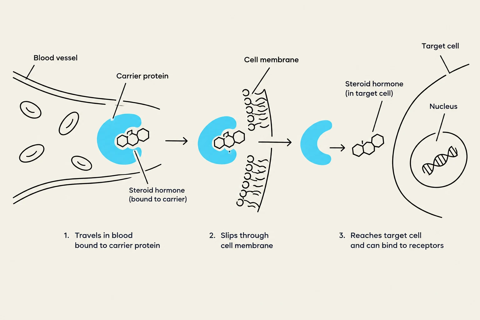
Lipophilic hormones, such as steroids (testosterone, estrogen, progesterone, cortisol) and thyroxine (an amine), do not dissolve well in water. Because blood is primarily water-based, these hormones cannot travel freely; instead, they must be bound to carrier proteins secreted into the blood. These proteins possess specific binding pockets that hold the hormone loosely, allowing it to dissociate and become "free" once it reaches its target tissue. Because they are lipophilic, these hormones can easily slip through the phospholipid bilayer of the plasma membrane to bind with intracellular receptors located inside the cell.
Exam Tip: Because lipophilic hormones must enter the cell and often trigger gene expression (switching genes on or off to make new proteins), their effects are generally slow to start but tend to be long-lasting.
Diagram illustrating how steroid hormones travel in the bloodstream bound to carrier proteins, pass through the cell membrane, and reach target cells to bind to receptors within the nucleus.
Hydrophilic Hormones (Water-loving)
Hydrophilic hormones, including peptides (insulin, glucagon, oxytocin, LH) and most neurotransmitters like epinephrine, dissolve easily in the blood and do not require carrier proteins. They circulate unbound and act by binding to receptors located on the plasma membrane (the cell surface).
Exam Tip: Because these hormones trigger a "second-messenger cascade" using proteins that already exist within the cell, their response is very fast (seconds to minutes) but typically short-lived.
Signaling Systems: Nervous vs. Endocrine
While both systems communicate, they differ significantly in speed and distance.
Nervous System: Provides rapid, localized communication via electrical impulses and hydrophilic neurotransmitters released at a synapse.
Endocrine System: Provides slower, body-wide signaling via the blood.
Neurohormones: These act as a bridge between the two systems. Neurohormones are peptide chemicals produced by specialized nerve cells but released into the bloodstream to regulate distant organs.
The Hypothalamus: The Master Relay Station
The hypothalamus is a structure of neural tissue that acts as the critical link between the nervous and endocrine systems. It regulates the pituitary gland through two distinct anatomical pathways: the posterior pituitary and the anterior pituitary.
The Posterior Pituitary (Neural Connection)
The hypothalamus is connected to the posterior pituitary via the hypothalamic-hypophyseal tract, which consists of neural tissue. Neurons in the hypothalamus produce peptide neurohormones, which travel down long axons to giant axon terminals located in the posterior pituitary. These terminals can store up to 30 days' worth of hormones.
Two key hormones are released here:
Oxytocin: Known as the "feel-good" or "bonding" hormone, it promotes uterine contractions during delivery, milk ejection during breastfeeding, and supports social bonding.
Anti-diuretic hormone (ADH): This hormone tells the kidneys to reabsorb water to conserve body fluids. A deficiency in ADH leads to frequent, watery urination, a condition known as Diabetes Insipidus.
The Anterior Pituitary (Circulatory Connection)
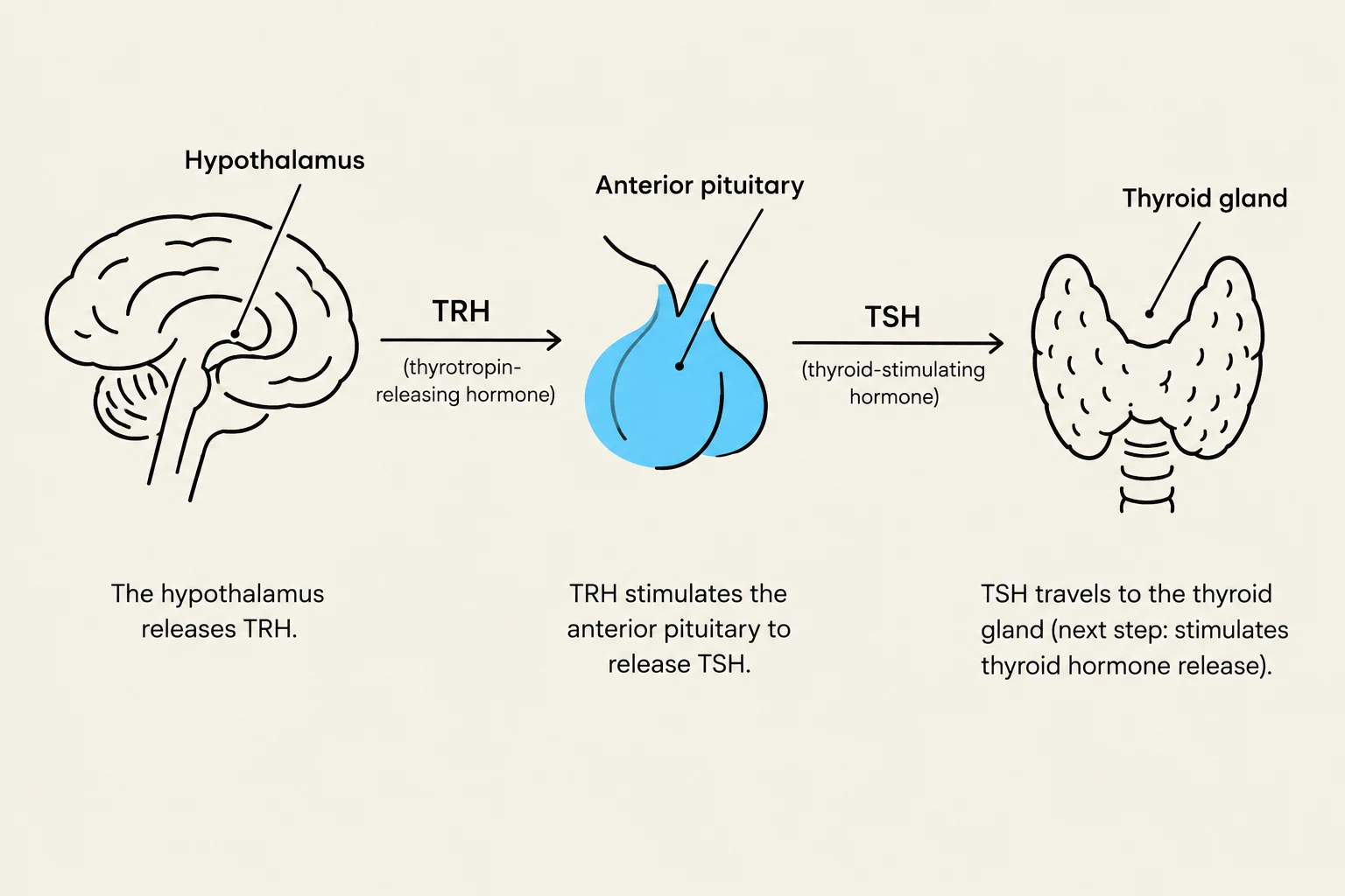
Unlike the posterior, the anterior pituitary is composed of glandular tissue. It is regulated by the hypothalamic-hypophyseal portal system, a specialized circulatory loop. Hypothalamic "releasing hormones" travel through this blood system to stimulate endocrine cells in the anterior pituitary to release their own specific hormones.
Hypothalamic Hormone
Anterior Pituitary Hormone
Target Tissue
GnRH
LH & FSH
Gonads (testes, ovaries)
TRH
TSH
Thyroid gland
CRH
ACTH
Adrenal glands
GHRH
Growth Hormone (GH)
Bone, muscle, liver, adipose
Dopamine (Inhibitory)
Prolactin
Mammary glands
The Thyroid Gland and Negative Feedback
The thyroid gland, located in the neck in front of the trachea, is a primary target of the anterior pituitary. It is composed of functional units called follicles. These follicles contain a "balloon-like" center filled with an oily substance called colloid, which stores thyroid hormones.
Thyroid Hormone Synthesis
Thyroid hormones are amines built from the amino acid tyrosine and iodine. The synthesis involves:
Follicular cells trapping iodide from the blood.
Combining iodide with tyrosine residues on a protein called thyroglobulin within the colloid.
Producing T4 (thyroxine), which is stable but inactive, and T3 (triiodothyronine), which is active but less stable.
Once TSH (Thyroid Stimulating Hormone) signals the gland, T4 and T3 are released into the blood. T4 is the primary product secreted, but target tissues must convert it into the active T3 to exert its effects.
Diagram illustrating the hormonal regulation of the thyroid gland, showing the roles of the hypothalamus, anterior pituitary, and thyroid gland in releasing TRH and TSH.
Function and Regulation
Thyroid hormones affect almost every nucleated cell in the body, primarily by increasing the basal metabolic rate (BMR) via the mitochondria. This increases heat production, oxygen consumption, and glucose use. They also impact heart rate, circulation, bone remodeling, and brain alertness.
To prevent hormone levels from becoming too high or too low, the body uses a negative feedback loop. When concentrations of T3 and T4 are high, they signal the hypothalamus to stop producing TRH and the anterior pituitary to stop producing TSH. As hormone levels drop, the inhibition is lifted, and the cycle begins again, maintaining homeostasis.
High [T3/T4]⟹↓TRH⟹↓TSH⟹↓Thyroid Hormone Production
Keep reading for free.
Create an account to finish this reading and make it part of your study routine.