An overview of the endocrine system, focusing on the hypothalamus and pituitary gland, their hormonal regulation, feedback mechanisms, and mechanisms of hormone action.
The endocrine system is a complex, integrated network of glands and organs that use chemical messengers, known as hormones, to maintain homeostasis—the stable internal environment required for survival. Unlike the nervous system, which uses electrical impulses for rapid communication, the endocrine system often manages slower, longer-lasting processes such as growth, metabolism, and reproduction. At the heart of this system lies the relationship between the hypothalamus and the pituitary gland, a partnership that allows the brain to translate neural signals into systemic hormonal responses.
The Hypothalamus: The Central Command Center
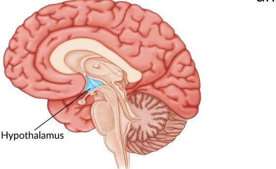
Located deep within the brain, below the thalamus and above the brainstem, the hypothalamus serves as the master regulator of the endocrine system. It acts as a bridge between the nervous and endocrine systems, integrating sensory information to coordinate autonomic, behavioral, and metabolic responses.
Diagram of the human brain highlighting the hypothalamus.
The hypothalamus is organized into distinct zones and nuclei that specialize in different homeostatic functions:
1. The Periventricular Zone
This thin strip lines the third ventricle and is the primary site for neuroendocrine regulation.
Paraventricular Nucleus (PVN): A master homeostatic nucleus that releases several key hormones, including Corticotropin-Releasing Hormone (CRH).
Arcuate Nucleus: Located near the base of the third ventricle, it is essential for regulating appetite, prolactin release, and monitoring adipose tissue.
Suprachiasmatic Nucleus (SCN): Positioned above the optic chiasm, it serves as the body's biological clock, synchronizing circadian rhythms with light-dark cycles via the retinohypothalamic tract.
2. The Medial Zone
This highly cellular region orchestrates autonomic and behavioral responses through several sub-regions:
Preoptic Area: Regulates temperature, sleep-wake cycles, and Gonadotropin-Releasing Hormone (GnRH).
Supraoptic Region: Contains the Supraoptic Nucleus (SON), which synthesizes Oxytocin (OT) and Antidiuretic Hormone (ADH/Vasopressin).
Tuberal Region: Includes the Ventromedial Nucleus (VMH) (the "satiety center") and the Dorsomedial Nucleus (DMH) (involved in energy expenditure and aggression).
Mammillary Region: Manages episodic memory and wakefulness via the tuberomammillary nucleus, the brain's sole source of histamine.
3. The Lateral Zone
Characterized by a more diffuse network of neurons, this zone is heavily involved in motivation and feeding:
Lateral Hypothalamic Nucleus: Known as the "feeding center".
Medial Forebrain Bundle (MFB): A massive neural highway that integrates reward, motivation, and pleasure.
The Pituitary Gland: The Master Gland
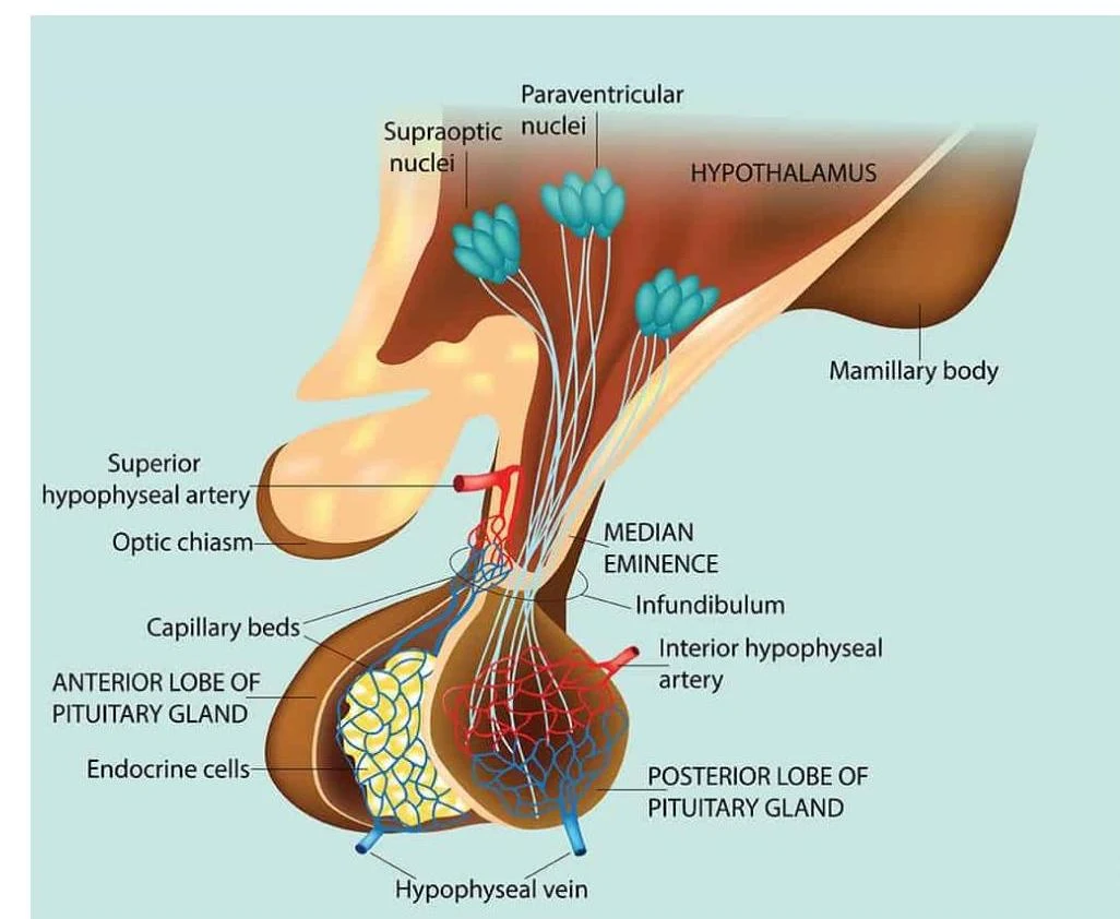
The pituitary gland is a pea-sized structure located in the sella turcica (a depression in the sphenoid bone). It is connected to the hypothalamus via the infundibulum (pituitary stalk). The gland is divided into two functionally and embryologically distinct lobes: the Anterior Pituitary (Adenohypophysis) and the Posterior Pituitary (Neurohypophysis).
The Anterior Pituitary (Adenohypophysis)
Derived from Ratke's pouch, the anterior pituitary is a true endocrine gland that produces and secretes its own hormones. However, its release is strictly controlled by "releasing" or "inhibiting" hormones from the hypothalamus.
These hypothalamic hormones travel to the anterior pituitary through a specialized capillary network called the hypophyseal portal system. This allows for rapid, concentrated communication between the brain and the gland.
Diagram illustrating the hypothalamus and pituitary gland, including key structures like the supraoptic and paraventricular nuclei, median eminence, infundibulum, mamillary body, and the anterior and posterior lobes of the pituitary gland with associated arteries and veins.
The anterior pituitary secretes several vital "tropic" hormones:
Hypothalamic Hormone
Anterior Pituitary Hormone
Target Organ
Primary Effect
GHRH (Growth Hormone-Releasing Hormone)
GH (Growth Hormone)
Liver, bone, muscle
Stimulates growth and metabolism
TRH (Thyrotropin-Releasing Hormone)
TSH (Thyroid-Stimulating Hormone)
Thyroid gland
Stimulates release of T3 and T4
CRH (Corticotropin-Releasing Hormone)
ACTH (Adrenocorticotropic Hormone)
Adrenal cortex
Stimulates steroidogenesis (cortisol)
GnRH (Gonadotropin-Releasing Hormone)
FSH & LH (Gonadotropins)
Gonads (testes/ovaries)
Regulates reproduction
Dopamine (Prolactin-Inhibiting Hormone)
PRL (Prolactin)
Mammary glands
Promotes milk production (inhibited by dopamine)
Key Hormone Deep Dives:
Growth Hormone (GH) Axis: GHRH stimulates the release of GH. GH then acts on the liver to produce Insulin-like Growth Factor 1 (IGF-1), which drives bone and muscle growth. This is regulated by Somatostatin, which acts as an inhibitor.
The HPA Axis (Stress Response): In response to stress, the hypothalamus releases CRH, which triggers the anterior pituitary to release ACTH. ACTH then travels to the adrenal cortex to stimulate the production of cortisol.
The Gonadotropins (FSH & LH):
In males, FSH stimulates spermatogenesis (sperm production) in the Sertoli cells, while LH stimulates the Leydig cells to produce testosterone.
In females, FSH promotes follicular development and estrogen secretion, while a massive "LH surge" triggers ovulation.
The Posterior Pituitary (Neurohypophysis)
Unlike the anterior lobe, the posterior pituitary is an extension of neural tissue. It does not produce hormones; instead, it stores and releases hormones that are synthesized in the hypothalamus (specifically the SON and PVN). These hormones travel down the hypothalamo-hypophyseal tract (a bundle of axons) to be released into the blood.
Antidiuretic Hormone (ADH / Vasopressin): Targets the kidneys to increase water reabsorption and constricts blood vessels to maintain blood pressure and electrolyte balance.
Oxytocin (OT): Targets the uterus to stimulate contractions during childbirth and the mammary glands to trigger milk ejection during breastfeeding.
Exam Tip: The "Production vs. Release" Distinction
Students often confuse the two lobes. Remember: The Anterior pituitary makes its own hormones (stimulated by the portal system), while the Posterior pituitary is just a storage warehouse for hormones made in the hypothalamus (delivered via neural axons).
Feedback Mechanisms and Homeostasis
The endocrine system maintains stability through biofeedback mechanisms. These loops ensure that once a physiological goal is met, the production of the hormone is dialed back.
Negative Feedback (The Stabilizer)
This is the most common regulatory mechanism. When the level of a target hormone (e.g., T3/T4 or Cortisol) rises in the blood, it acts on the hypothalamus and/or the anterior pituitary to inhibit the release of the "upstream" stimulating hormones.
Example: The HPA Axis
Stress → Hypothalamus releases CRH.
CRH → Anterior Pituitary releases ACTH.
ACTH → Adrenal Cortex releases Cortisol.
High Cortisol→ Signals Hypothalamus and Pituitary to stop releasing CRH and ACTH.
Positive Feedback (The Amplifier)
This is rare and used for processes that need to reach a climax or "explosive" conclusion. Instead of shutting the system down, the response increases the stimulus.
Example: Childbirth
The baby's head pushes against the cervix.
Nerve impulses signal the hypothalamus to release Oxytocin.
Oxytocin causes uterine contractions.
Contractions push the baby harder against the cervix, triggering even more oxytocin.
Mechanisms of Hormone Action
Hormones cannot work unless they find a specific receptor on or in a target cell. The mechanism of action depends on whether the hormone is water-soluble or lipid-soluble.
Hydrophilic (water-soluble) hormones, such as peptide hormones (insulin, GH, TSH) and catecholamines, cannot pass through the fatty cell membrane.
Process: The hormone (the "first messenger") binds to a receptor on the cell surface. This activates an internal enzyme (like adenylate cyclase), which converts ATP into cyclic AMP (cAMP).
Result: cAMP acts as a second messenger, triggering a cascade of protein kinases that alter cellular metabolism rapidly.
Lipophilic (lipid-soluble) hormones, such as steroid hormones (cortisol, testosterone, estrogen) and thyroid hormones (T3, T4), can diffuse directly through the plasma membrane.
Process: Once inside, they bind to receptors in the cytoplasm or nucleus.
Result: The hormone-receptor complex binds directly to the cell's DNA, acting as a transcription factor to turn specific genes "on" or "off," leading to the synthesis of new proteins.
Clinical Applications and Integration
Disruptions in these feedback loops lead to significant clinical disorders.
Thyroid Disorders:
Hyperthyroidism (Thyrotoxicosis): Excess T3/T4 leads to a "sped up" metabolism, causing tachycardia, weight loss, and heat intolerance.
Hypothyroidism (Myxedema): A deficiency leads to a "slowed down" metabolism, causing bradycardia, weight gain, and fatigue.
Adrenal Disorders:
Cushing's Syndrome: Caused by chronic cortisol excess, presenting with "moon face," central obesity, and a "buffalo hump".
Addison's Disease: Caused by adrenal insufficiency (lack of mineralocorticoids/cortisol), leading to severe hypotension and electrolyte imbalance.
Glucose Regulation:
Type 1 Diabetes: Absolute insulin deficiency due to beta-cell destruction.
Type 2 Diabetes: Characterized by insulin resistance at the receptor level.
Clinical Reasoning Tip: Primary vs. Secondary Disorders
When diagnosing endocrine issues, look at the feedback loop. If a patient has low thyroid hormone (T3/T4) but high TSH, the problem is primary (the thyroid gland itself is failing). If both T3/T4 and TSH are low, the problem is secondary (the pituitary gland is failing to send the signal).
Keep reading for free.
Create an account to finish this reading and make it part of your study routine.