


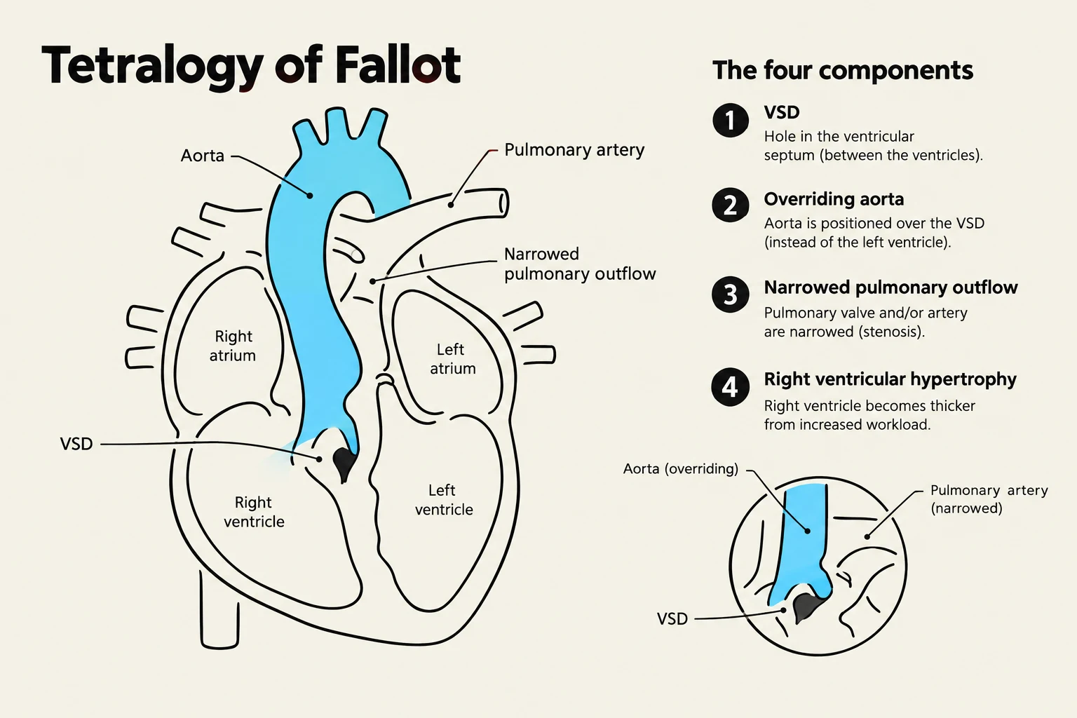
The heart is the central engine of the cardiovascular system, responsible for distributing oxygen, nutrients, and hormones to tissues while removing metabolic waste products like and nitrogenous wastes. Understanding how this complex pump forms during gestation and how its circulation shifts at birth is fundamental to clinical medicine, particularly when diagnosing congenital malformations.
Embryological Development: From Tube to Chambers
The journey of the heart begins during the second week of gestation with the formation of angioblasts. By the end of this period, two separate endocardial tubes fuse to form a single, primitive heart tube. This primitive tube is composed of three distinct layers: the inner endocardium, the muscular myocardium, and the outer epicardium (also known as the visceral pericardium).
As development progresses into the fourth week, the heart undergoes a dramatic transformation known as looping or deformation. The primitive tube does not simply grow; it bends and rotates. The atrium and the sinus venosus migrate cranially (upward), while the ventricle rotates ventrally (forward). This complex movement is essential to position the chambers correctly for the eventual separation of systemic and pulmonary circulations.
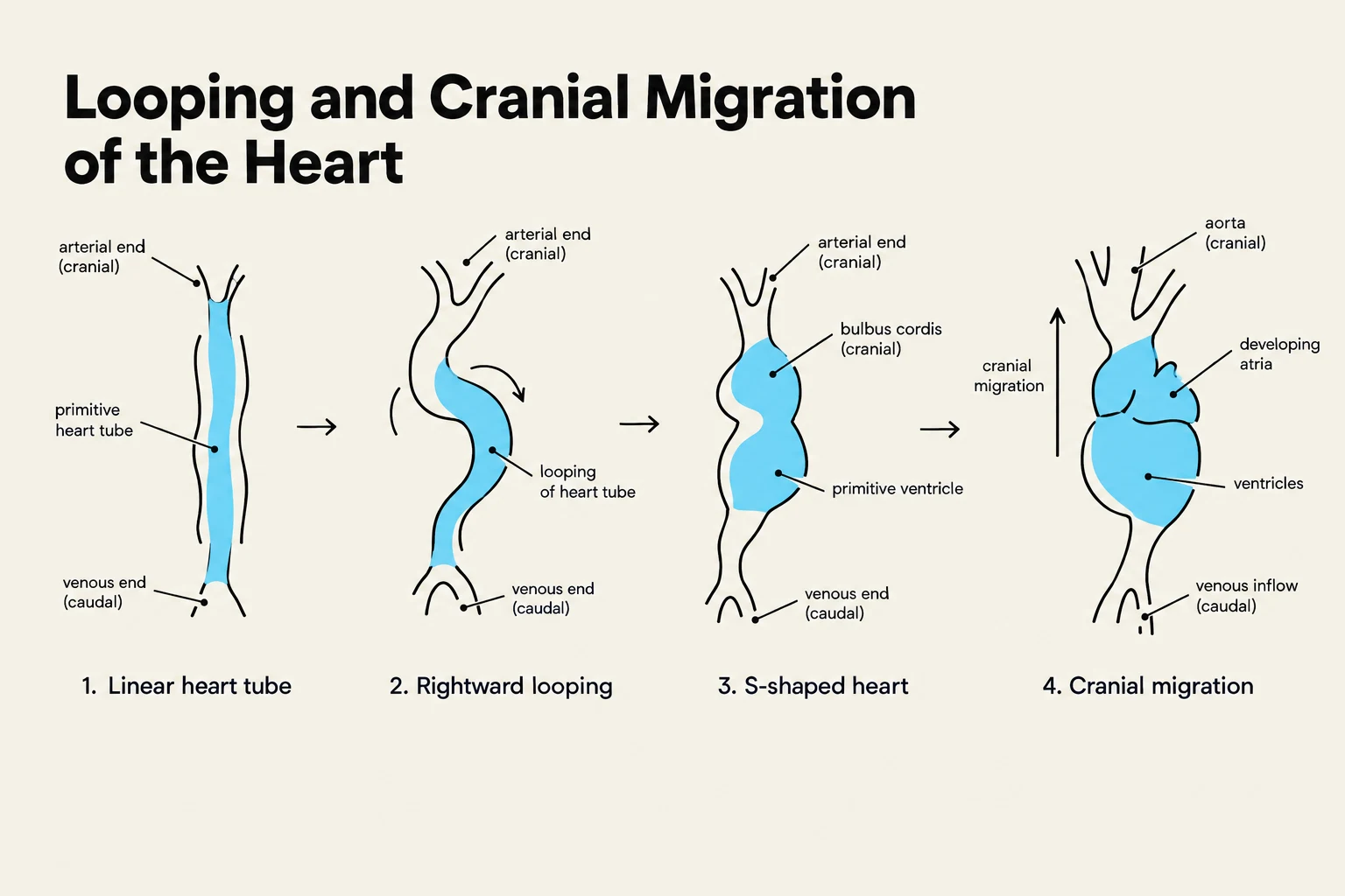
Atrial and Ventricular Septation
To transition from a single tube to a four-chambered organ, the heart must develop internal walls, or septa.
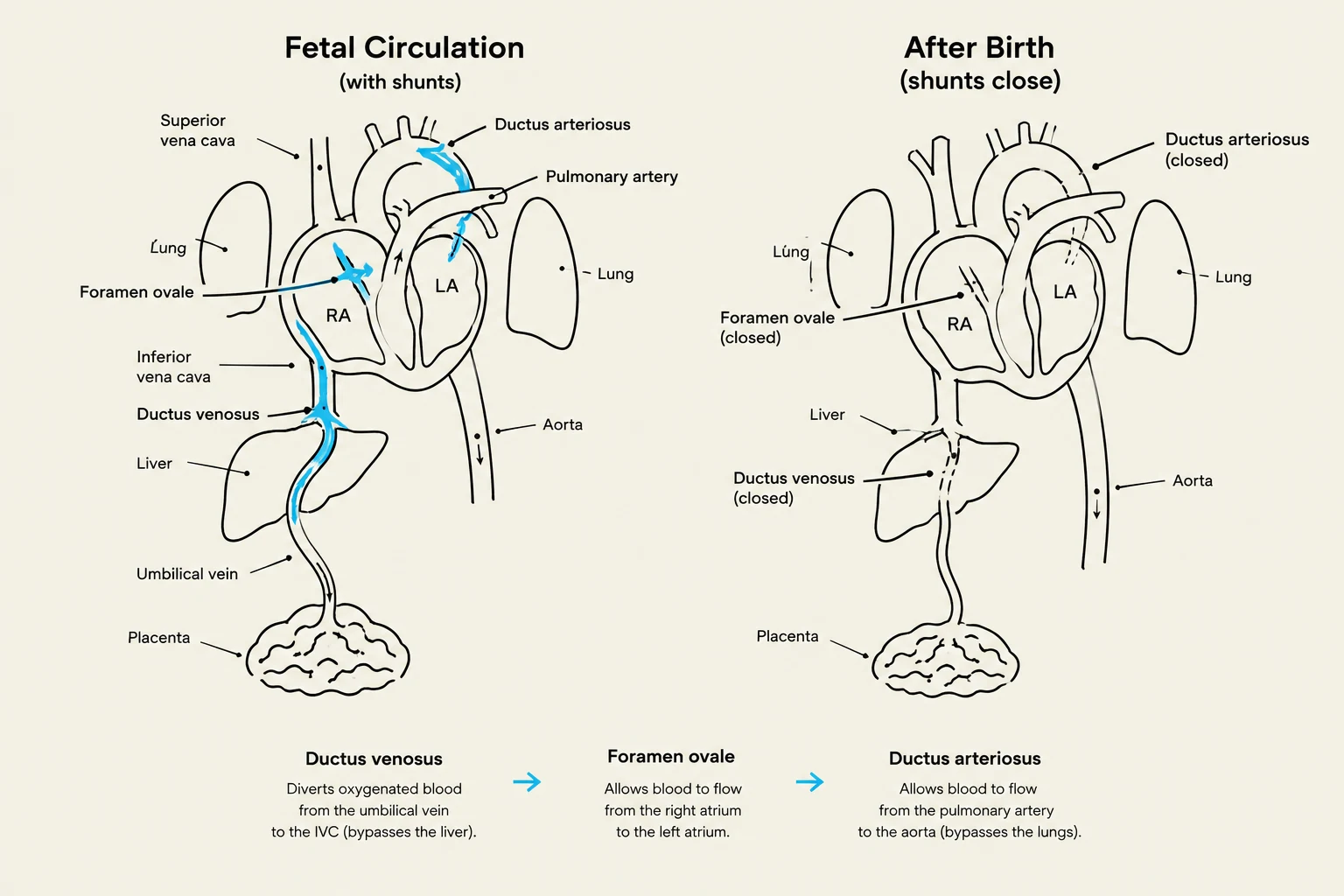
- Atrial Septation: The separation of the left and right atria occurs via the septum primum and the septum secundum. During this process, an opening called the foramen ovale (specifically the ostium secundum) is formed. This opening is a critical physiological feature in the fetus, allowing blood to flow from the right atrium directly to the left atrium, thereby bypassing the non-functional, fluid-filled lungs.
- Ventricular Septation: The ventricles are divided by the interventricular septum, which consists of a muscular part and a membranous part.
- Great Vessel Separation: The truncus arteriosus and the bulbus cordis are eventually divided into the aorta and the pulmonary trunk by spiraling ridges that revolve around each other.
Keep reading for free.
Create an account to finish this reading and make it part of your study routine.
Continue reading freeAlready have an account? Log in
Updated