Explains the electron transport chain (ETC), its role in cellular respiration, and the function of various electron carriers like FMN, Fe-S clusters, Coenzyme Q, and cytochromes in generating ATP. It also covers redox potential and the production of reactive oxygen species.
The survival of aerobic organisms depends on the ability to convert the chemical energy stored in nutrients into a form that cells can use: ATP. This conversion is achieved through the Electron Transport Chain (ETC), a series of protein complexes and mobile carriers embedded within the inner mitochondrial membrane (IMM). As electrons move through this chain, they undergo a series of oxidation-reduction (redox) reactions, releasing free energy that is used to pump protons across the membrane, creating an electrochemical gradient.
The Principles of Electron Transfer
To understand how electrons move through the ETC, we must first understand the concept of redox potential (E0′). A molecule's reduction potential is a measure of its tendency to accept electrons.
Reducing agents (like NADH) have a negative E0′ and tend to donate electrons.
Oxidizing agents (like O2) have a positive E0′ and tend to accept electrons.
In the ETC, electrons flow "downhill" from carriers with low reduction potential to carriers with high reduction potential. This spontaneous flow is coupled to the "uphill" movement of protons from the mitochondrial matrix to the intermembrane space. The relationship between the change in reduction potential (ΔE0′) and the standard free energy change (ΔG∘′) is defined by the following equation:
ΔG∘′=−nFΔE0′
Where n is the number of electrons transferred and F is the Faraday constant.
Exam Tip: Remember that a larger, positive ΔE0′ results in a more negative ΔG∘′, meaning more energy is released during the electron transfer. This energy is what drives the proton pumps!
Specialized Electron Carriers
The transfer of electrons from NADH to O2 is not a single jump; it is an indirect process facilitated by four large protein complexes and several specialized prosthetic groups and mobile carriers.
Flavin Mononucleotide (FMN)
Within Complex I (NADH-Q oxidoreductase), the first recipient of electrons from NADH is Flavin mononucleotide (FMN). FMN is a derivative similar to FAD but lacks the large nucleotide component. It acts as a crucial bridge, accepting two electrons from NADH to initiate the chain.
Iron-Sulfur (Fe-S) Clusters
As electrons move through the large protein complexes, they are passed along Iron-sulfur (Fe-S) clusters. These are non-heme clusters consisting of iron associated with sulfur within the protein structure. They are highly efficient at undergoing the oxidation-reduction reactions necessary to shuttle electrons between different parts of the complexes.
Coenzyme Q (Ubiquinone)
Unlike the protein-bound clusters, Coenzyme Q (Ubiquinone, Q) is a highly important, mostly hydrophobic mobile electron carrier. Because it is hydrophobic, it resides within the lipid bilayer of the inner mitochondrial membrane, forming what is known as the Q-pool.
Coenzyme Q is unique because it can exist in multiple oxidation states and can bind both protons and electrons. This allows it to shuttle electrons from both Complex I and Complex II to Complex III.
Diagram illustrating the movement of Coenzyme Q within the inner mitochondrial membrane, between Complex III and Complex IV.
Cytochromes and Heme Groups
The final stages of the ETC involve cytochromes, which are proteins containing heme prosthetic groups. While heme groups are famous for their roles in myoglobin and hemoglobin, their function in the ETC is distinct.
In hemoglobin, the iron remains in a stable oxidation state to bind oxygen. However, in the heme groups of cytochromes, the iron actively participates in electron transfer by cycling between the reduced Fe2+ state and the oxidized Fe3+ state. These cytochromes are found in Complex III and Complex IV, facilitating the final movement of electrons toward oxygen.
Summary of Electron Flow
The path electrons take depends on their initial donor.
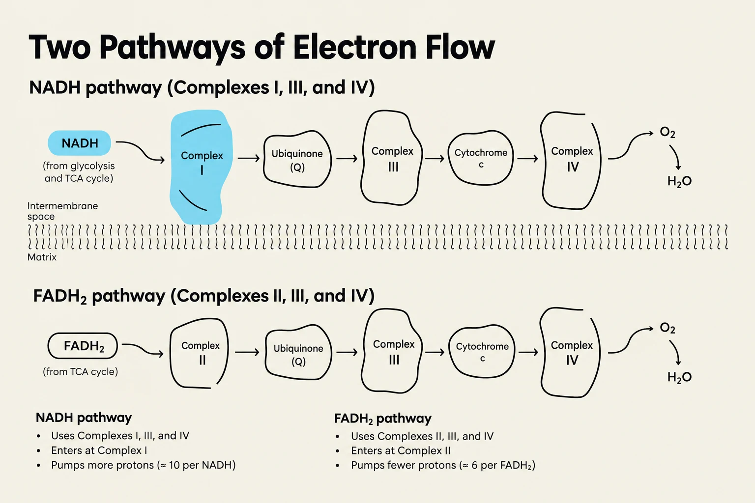
Starting with NADH: Electrons enter at Complex I → Coenzyme Q → Complex III → Cytochrome c → Complex IV →O2. This pathway results in the pumping of 10 protons across the membrane.
Starting with FADH2: Electrons enter at Complex II (Succinate-Q reductase) → Coenzyme Q → Complex III → Cytochrome c → Complex IV →O2. Because Complex II does not pump protons, this pathway only results in the pumping of 6 protons.
Study Question: Why does NADH lead to more ATP generation than FADH2?
Answer: Because NADH enters the chain at Complex I, it triggers the pumping of more protons (10 vs 6) into the intermembrane space. A larger proton gradient provides more potential energy for ATP synthase to produce ATP.
Diagram illustrating the two pathways of electron flow in cellular respiration: the NADH pathway and the FADH2 pathway, detailing the complexes involved and their respective proton pumping efficiencies.
The Danger of Incomplete Reduction
While the ETC is highly efficient, it is not perfect. Occasionally, oxygen undergoes incomplete reduction, leading to the production of Reactive Oxygen Species (ROS), such as superoxide ions and peroxide. These molecules can damage DNA, proteins, and membranes. To combat this, cells utilize specialized enzymes like superoxide dismutase and catalase to neutralize these threats.
Keep reading for free.
Create an account to finish this reading and make it part of your study routine.