Efferent Division: Autonomic and Somatic Motor Control
Explains the efferent division of the peripheral nervous system, detailing the somatic motor system for voluntary control and the autonomic nervous system (ANS) for involuntary functions. Covers the sympathetic ('fight or flight') and parasympathetic ('rest and digest') branches, their neurotransmitters, and central co
The peripheral nervous system is divided into sensory (afferent) and motor (efferent) divisions. While the afferent division carries information toward the central nervous system (CNS), the efferent division is responsible for carrying motor commands away from the CNS to effectors like muscles and glands. This motor control is further subdivided into two distinct systems: the somatic nervous system, which governs voluntary movement, and the autonomic nervous system (ANS), which manages involuntary physiological processes.
Somatic Motor Control
The somatic nervous system provides voluntary control over skeletal muscles. Unlike the autonomic system, which uses a two-neuron chain, the somatic division typically consists of a single, heavily myelinated motor neuron that originates in the CNS and extends its axon all the way to the target muscle.
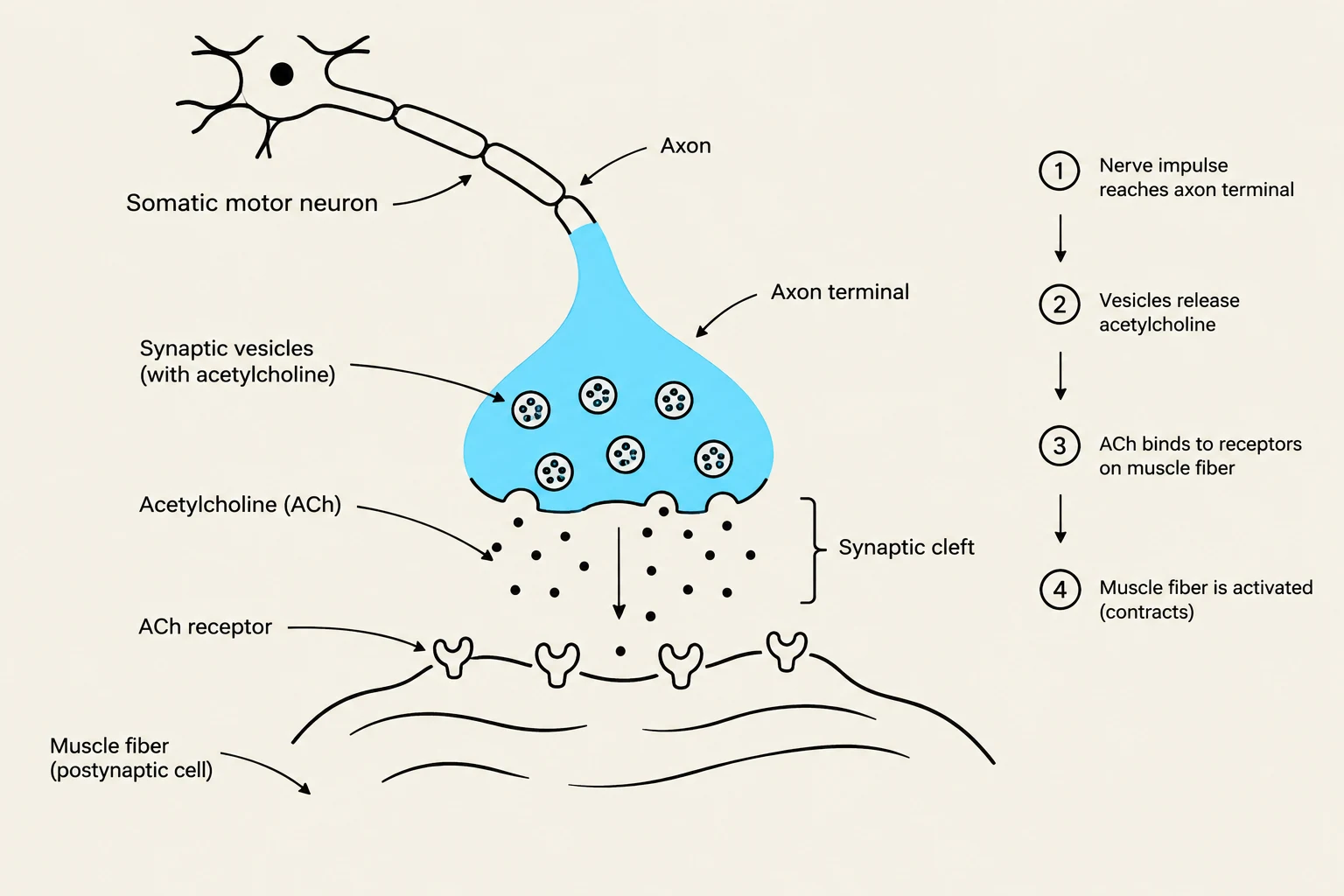
The point of communication between the motor neuron and the muscle fiber is called the neuromuscular junction. At this synapse, the somatic motor neuron releases the neurotransmitter acetylcholine (ACh) into the synaptic cleft. The ACh molecules bind to specialized nicotinic ACh receptors located on the motor end plate of the muscle fiber. This binding triggers an influx of calcium ions (Ca2+), which leads to an action potential in the muscle fiber and subsequent muscle contraction.
Diagram illustrating the process of neuromuscular transmission, where a somatic motor neuron communicates with a muscle fiber via a synapse. It shows the release of acetylcholine from synaptic vesicles, its binding to receptors on the muscle fiber, and the subsequent activation and contraction of the muscle fiber.
The Autonomic Nervous System (ANS)
The primary goal of the autonomic nervous system is to maintain homeostasis—the stable internal environment required for survival. To achieve this, the ANS regulates involuntary functions such as heart rate, digestion, and respiratory rate.
The ANS operates through a unique two-neuron pathway. A preganglionic neuron exits the CNS and travels to an autonomic ganglion (a cluster of nerve cell bodies outside the CNS), where it synapses with a postganglionic neuron. This second neuron then extends to the target tissue.
The Sympathetic Division: "Fight or Flight"
The sympathetic division prepares the body for physical activity or emergency situations. When activated, it stimulates responses that maximize energy availability and blood flow to vital organs, such as:
Pupil dilation
Increased heart rate and blood volume
Bronchiole dilation to increase airflow
Fat breakdown for energy
Conversely, the sympathetic branch inhibits non-essential functions during a crisis, such as digestion, pancreas secretion, and urination.
A specialized component of this system is the adrenal medulla. This structure acts as a modified sympathetic ganglion where chromaffin cells (modified postganglionic neurons) release epinephrine directly into the bloodstream. Because epinephrine enters the blood, it acts as a neurohormone, allowing the "fight or flight" signal to reach tissues throughout the entire body.
The Parasympathetic Division: "Rest and Digest"
The parasympathetic division opposes the sympathetic branch to conserve energy and promote maintenance functions. It is often referred to as the "rest and digest" system. Its primary actions include:
Constricting pupils and bronchioles
Slowing the heart rate
Stimulating digestion and insulin release
Promoting urination
Exam Tip: Remember that the two branches are antagonistic. They often have opposite effects on the same organ to maintain a fine balance of physiological function.
Neurotransmitters and Receptor Mechanisms
The way the ANS communicates with target cells depends heavily on the specific neurotransmitters used and the receptors they bind to.
Sympathetic Neurotransmission
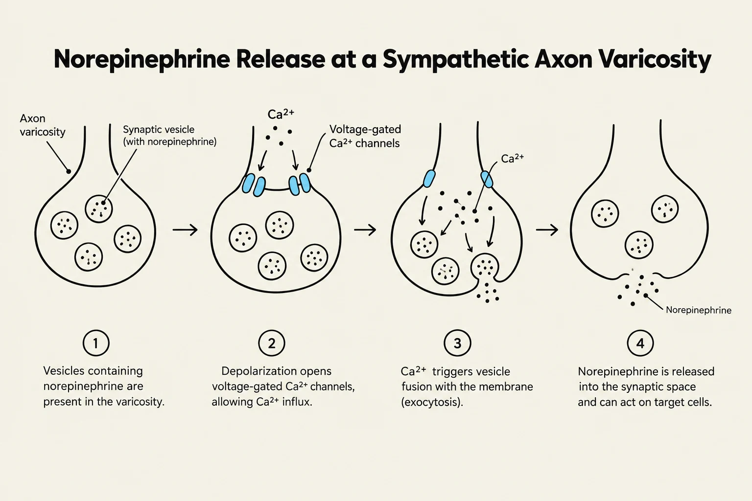
In the sympathetic division, the postganglionic neurons typically release norepinephrine (NE) at the target tissue. The release of NE follows a specific sequence:
An action potential arrives at the axon varicosity (a swelling along the axon).
Depolarization opens voltage-gated Ca2+ channels.
The influx of Ca2+ triggers the exocytosis of synaptic vesicles containing NE.
NE binds to adrenergic receptors on the target cell.
Once the signal is sent, NE must be removed from the synapse to prevent continuous stimulation. This occurs through diffusion, reuptake into the neuron, or metabolism by the enzyme monoamine oxidase (MAO).
Parasympathetic Neurotransmission
In contrast, the parasympathetic division primarily utilizes acetylcholine (ACh) to communicate with its target organs.
Diagram illustrating the process of norepinephrine release at a sympathetic axon varicosity, involving vesicle fusion and exocytosis triggered by calcium influx.
Autonomic Control Centers
While the neurons of the ANS carry out the "work," the coordination of these signals happens in the brain. Several autonomic control centers integrate sensory information and send out instructions to maintain homeostasis:
Hypothalamus: Acts as a master regulator for temperature control, water balance, and eating behavior.
Medulla Oblongata: Controls vital life-sustaining functions, including cardiac output (heart rate), blood pressure, respiration, swallowing, and vomiting.
Pons: Works alongside the medulla to regulate respiratory rhythms.
In clinical and research settings, scientists use autonomic agonists and antagonists—substances that either mimic or block the effects of these neurotransmitters—to study and manipulate these complex pathways.
Keep reading for free.
Create an account to finish this reading and make it part of your study routine.