Effects of Temperature and Organic Solvents on Cellular Membranes
This reading explores how environmental stressors like temperature fluctuations and organic solvents impact the integrity of cellular membranes. It uses beet cells and the betacyanin pigment as a model to demonstrate membrane damage and discusses spectrophotometry as a method for quantifying this damage.
In the study of biology, understanding how cells maintain their internal environment is fundamental. At the heart of this maintenance are cellular membranes—thin, delicate structures that act as gatekeepers, regulating what enters and exits the cell. While these membranes are incredibly efficient at organizing life processes, they are also highly susceptible to environmental stressors. By using the beet (Beta vulgaris) as a biological model, researchers can visually observe how physical and chemical stresses compromise these vital barriers.
The Architecture of the Cell: Barriers and Pigments
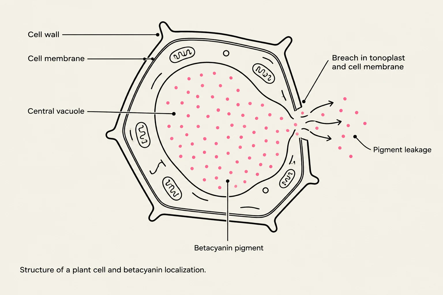
Diagram illustrating the structure of a plant cell and the localization of betacyanin pigment, showing pigment leakage due to a breach in the tonoplast and cell membrane.
To understand how stress damages a cell, we must first understand the layers that protect it. A plant cell, such as those found in a beet, is protected by several distinct layers:
The Cell Wall: The outermost rigid layer that provides structural support.
The Cell Membrane: A thin, selectively permeable barrier located inside the cell wall.
The Vacuole: A large central organelle that stores various substances.
The Tonoplast: The specific membrane that surrounds the vacuole, separating its contents from the rest of the cytoplasm.
The beet is an ideal experimental system because of a specific molecule called betacyanin. This is a reddish pigment that is localized almost entirely within the large central vacuoles. In a healthy, intact cell, the betacyanin is trapped behind both the tonoplast and the cell membrane. However, if these membranes are damaged, the pigment will undergo diffusion, leaking out of the cell and into the surrounding water. The intensity of the red color in the surrounding liquid serves as a direct indicator of how much membrane damage has occurred.
The Fluid Mosaic Model: The Role of Phospholipids
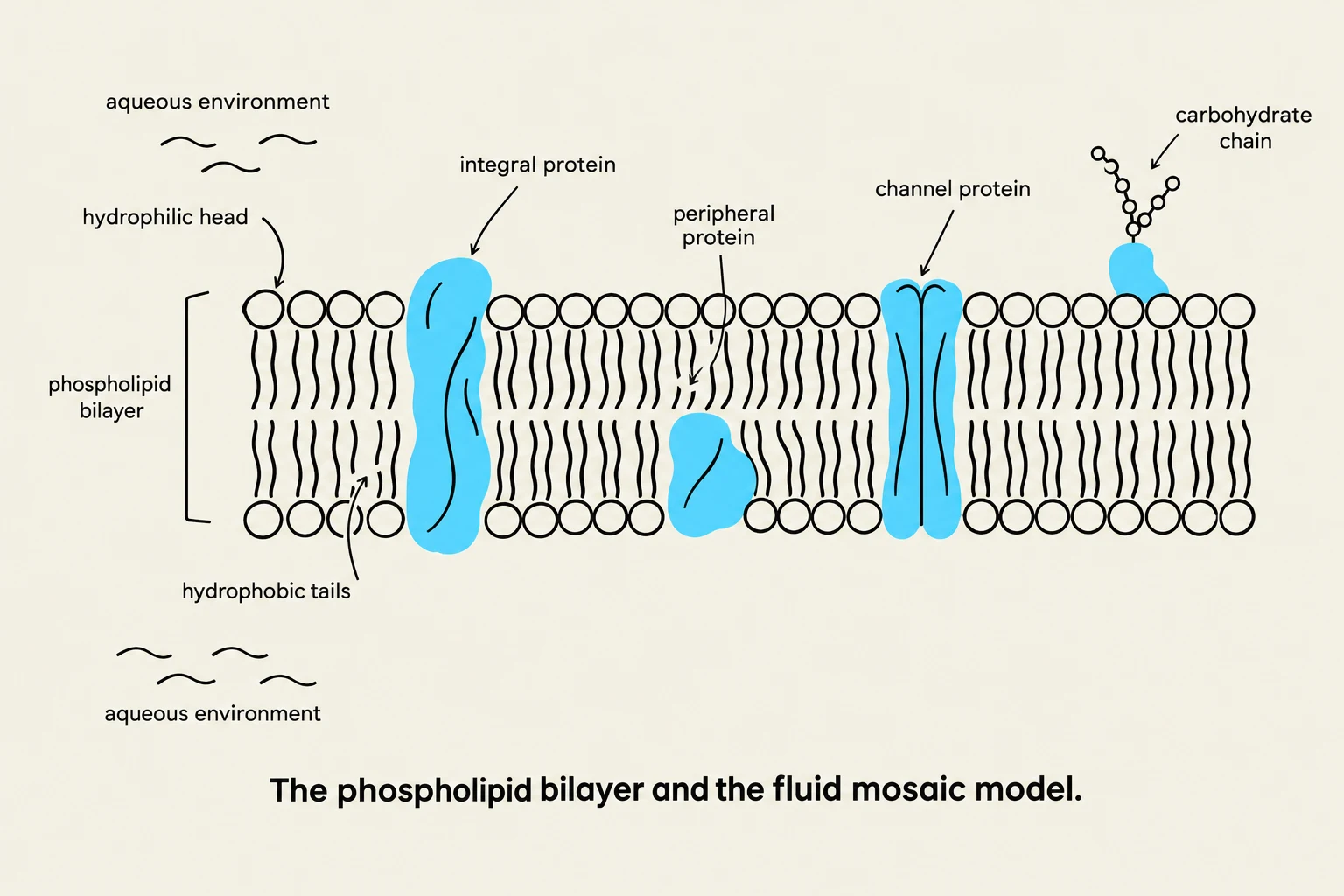
Diagram illustrating the phospholipid bilayer and the fluid mosaic model, showing integral and peripheral proteins, and a carbohydrate chain.
The integrity of the cell membrane relies on its unique molecular structure. Membranes are composed of a phospholipid bilayer. A phospholipid is an amphipathic molecule, meaning it possesses both hydrophilic ("water-loving") and hydrophobic ("water-fearing") regions.
The Hydrophilic Head: Consists of a polar phosphate group and a glycerol chain that seeks contact with water.
The Hydrophobic Tail: Consists of two nonpolar fatty acid chains that avoid water.
Because of these dual affinities, phospholipids naturally self-assemble into a double-layered sheet where the hydrophobic tails face inward, forming a protected core, while the hydrophilic heads line the surfaces in contact with the aqueous environment. This bilayer is embedded with various proteins that facilitate transport and signal detection, creating what is known as the fluid mosaic model. This elegant assembly is stable and self-repairing, but it is also vulnerable to specific types of stress.
Exam Tip: Selective Permeability
Remember that the membrane is "selectively permeable." This means it does not allow everything to pass through. Generally, small, uncharged, nonpolar, lipid-soluble molecules pass through the lipid core most easily, while polar or charged molecules often require specialized protein channels.
Temperature Stress: Heat and Ice
Temperature fluctuations can disrupt the membrane through two very different physical mechanisms:
High Temperatures
Extreme heat increases the kinetic energy of the molecules within the membrane. This leads to violent molecular collisions that can physically degrade the membrane's ability to function as a stable barrier to diffusion.
Freezing Temperatures
When temperatures drop below freezing, water inside and around the cell begins to crystallize into ice. Because of the specific alignment of hydrogen bonds, ice expands as it forms. This expansion can create physical pressure that ruptures the cell membrane and the tonoplast, causing the contents to spill out.
Organic Solvent Stress: Dissolving the Barrier
While temperature acts through kinetic energy and physical expansion, organic solvents like acetone and methanol attack the membrane chemically.
The core of the membrane is held together by the hydrophobic interactions of the lipid tails. Organic solvents are capable of dissolving these lipids. When the solvent interacts with the phospholipid bilayer, it disrupts the cohesive forces holding the membrane together, effectively dissolving the barrier. Once the lipid bilayer is dissolved, the betacyanin pigment is no longer contained and leaks into the external solution.
Quantifying Damage
In a laboratory setting, the extent of membrane injury is often quantified by observing the color intensity of the solution. Researchers may use a visual scale (e.g., 0 for colorless to 10 for darkest red) or more precise scientific instruments.
Using Spectrophotometry
This diagram illustrates the relationship between pigment concentration and light absorbance, showing how increasing concentration leads to higher absorbance.
To move beyond visual estimation, scientists use a spectrophotometer. This instrument measures how much light a solution absorbs or transmits. Every chemical has a unique "fingerprint" of wavelengths it absorbs.
By directing a specific wavelength of light through a sample of beet juice, the spectrophotometer can determine the concentration of betacyanin based on two key metrics:
Transmittance: The amount of light that passes through the sample. As the concentration of pigment increases, transmittance decreases.
Absorbance: The amount of light retained by the sample. As the concentration of pigment increases, absorbance increases.
The relationship between absorbance and concentration is governed by the Beer-Lambert Law, which states that absorbance is directly proportional to the concentration of the substance in the solution. This allows researchers to create a standard curve—a graph of known concentrations versus their absorbance—to accurately calculate the exact amount of pigment leaked by a treated beet cell.
Keep reading for free.
Create an account to finish this reading and make it part of your study routine.