Explores the Developmental Origins of Health and Disease (DOHaD) framework, focusing on how early life environmental stimuli can lead to lasting physiological changes through epigenetic programming, including DNA methylation and histone modification.
The concept of health and disease is no longer viewed solely through the lens of adult lifestyle or genetic inheritance. Instead, modern science recognizes a profound connection between early life environments and long-term physiological outcomes. This connection is encapsulated in the framework of the Developmental Origins of Health and Disease (DOHaD), which posits that specific stimuli or "insults" during critical developmental windows can leave lasting effects on the structure and function of the body.
Understanding DOHaD and Developmental Programming
Developmental programming occurs when an environmental stimulus—such as nutrition, stress, or toxins—acts during a critical window of development. This can lead to permanent changes in organ structure, cellular aging, or the establishment of altered homeostatic setpoints. For example, a prenatal insult might lead to changes in kidney structure that increase the risk of hypertension later in life.
These changes are often the result of Gene-Environment (GXE) interactions. While an individual's genotype provides the blueprint, the environment determines how those genes are expressed. Different genotypes may respond to the same environmental stimulus in divergent ways, leading to a wide variety of phenotypes from the same starting genetic code.
Exam Tip: Remember that DOHaD is not just about "damage." It is about the body's attempt to adapt to its environment, which can result in "mismatched" physiological setpoints if the adult environment differs from the prenatal one.
The Role of Epigenetic Programming
If DNA is the hardware of life, epigenetics is the software that determines which programs are running. Epigenetics is the study of changes in gene expression that do not involve alterations to the underlying DNA sequence itself. Instead, these changes involve chemical modifications to the DNA or the proteins (histones) around which DNA is wrapped.
These modifications create a chemical "signature" on the genes, determining whether they are "switched on" (active) or "switched off" (silent). This process allows cells with identical DNA to differentiate into specialized types, such as neurons or muscle cells.
Major Epigenetic Mechanisms
There are three primary mechanisms through which epigenetic programming occurs:
DNA Methylation: This involves the addition of methyl groups to DNA, typically at CpG sites (where a cytosine is followed by a guanine). When methyl groups are added to a gene's promoter or enhancer regions, they prevent RNA polymerase from binding, effectively silencing the gene.
Histone Modification: DNA is wrapped around proteins called histones to form chromatin. Chemical modifications to the "tails" of these histones—such as acetylation or methylation—change how tightly the DNA is packed.
Acetylation: Generally associated with "open" chromatin (euchromatin), making genes accessible for transcription.
Methylation: Can lead to "condensed" chromatin (heterochromatin), which silences gene expression.
Non-Coding RNAs (ncRNAs): Molecules like microRNAs (miRNAs) and long non-coding RNAs (lncRNAs) can regulate gene expression post-transcriptionally by promoting the degradation of messenger RNA (mRNA) or inhibiting its translation.
Maternal Behavior and the Hippocampus
One of the most striking examples of epigenetic programming involves maternal care and its impact on the brain's stress response system. In animal models, maternal behavior—specifically tactile stimulus (such as licking and grooming)—serves as a powerful environmental signal that shapes the offspring's epigenome.
The HPA Axis and the Glucocorticoid Receptor
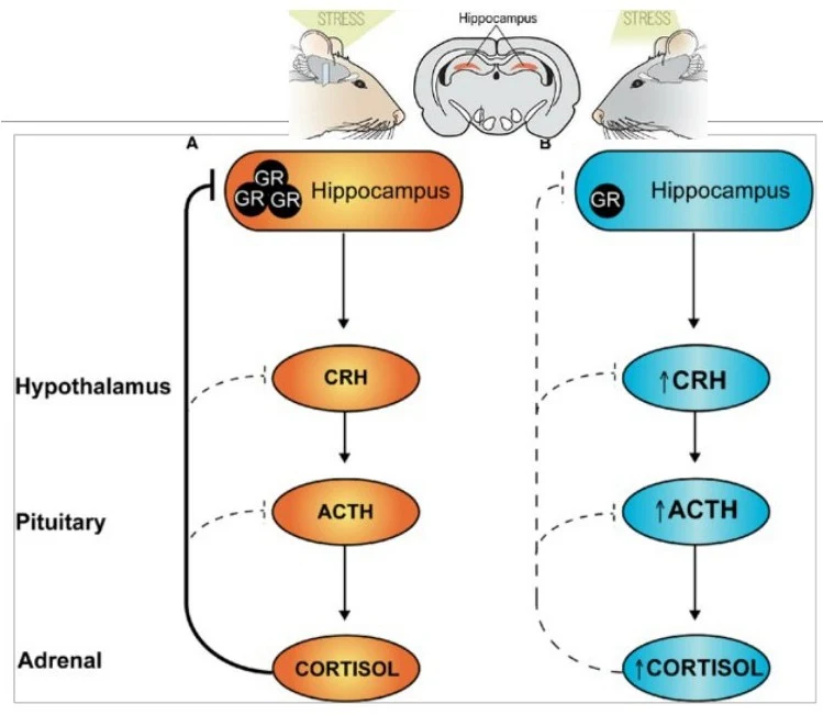
The Hypothalamic-Pituitary-Adrenal (HPA) axis is the body's central stress response system. A key component in regulating this system is the Glucocorticoid Receptor (GR), located in the hippocampus. The hippocampus provides negative feedback to the HPA axis; when GRs detect cortisol (or corticosterone in rats), they signal the system to shut down the stress response.
Research has shown that maternal care directly regulates the expression of the GR gene in the hippocampus through the following mechanism:
Tactile Stimulus: High levels of maternal grooming/licking provide sensory input to the pup.
Transcription Factor Binding: This stimulus promotes the binding of the transcription factor NGFI-A to the Glucocorticoid Receptor (GR) gene promoter.
Epigenetic Remodeling: Increased NGFI-A binding is associated with decreased DNA methylation and increased histone acetylation at the promoter site.
Increased Expression: Because the promoter is unmethylated and the chromatin is "open," the GR gene is highly expressed.
This illustration depicts the effect of stress on gene methylation in the hippocampus of mice, showing differences between genes rarely methylated and genes highly methylated.
This diagram illustrates the hypothalamic-pituitary-adrenal (HPA) axis, showing the regulation of cortisol release in response to stress. The left side (A) depicts negative feedback from the hippocampus on cortisol production, while the right side (B) shows the stimulatory effect of stress, leading to increased CRH, ACTH, and cortisol.
As a result, pups receiving high levels of maternal care develop a more efficient HPA axis with robust negative feedback, making them more resilient to stress in adulthood. Conversely, low maternal care leads to hypermethylation of the GR promoter, fewer receptors, and a hyperreactive stress response.
Key Concept: The link between maternal behavior and the HPA axis is a classic example of how an environmental "experience" is converted into a stable "epigenetic signature".
Summary of DOHaD Mechanisms
To summarize the ways in which early life environments impact long-term health, we can look at the four primary pillars of DOHaD mechanisms:
Mechanism
Description
Epigenetics
Chemical modifications (DNA methylation, histone changes) that alter gene expression.
Altered Organ Structure
Physical changes in the development of organs (e.g., "brain sparing" during hypoxia).
Cellular Aging
Changes in telomere length and rate of attrition, affecting lifespan and disease susceptibility.
Altered Homeostatic Setpoints
Changes in physiological regulation, such as HPA axis hyperreactivity or altered metabolic rates.
Understanding these mechanisms is vital for developing early interventions. Since the epigenome is plastic during critical windows, education, nutritional support, and social interventions can potentially mitigate the risks associated with adverse early-life environments.
Keep reading for free.
Create an account to finish this reading and make it part of your study routine.