Explores the molecular structure of DNA, including nucleotide composition, the double helix, complementary base pairing, and supercoiling. Details the semi-conservative replication process, key enzymes involved, and the synthesis of leading and lagging strands.
The ultimate goal of a cell is to duplicate its genetic information and pass it accurately to the next generation. This process is governed by the precise molecular structure of Deoxyribonucleic Acid (DNA), a molecule that serves as the blueprint for all cellular functions. Understanding how this information is stored in the chemical sequence of nucleotides and how it is copied through complex enzymatic machinery is fundamental to the study of molecular biology.
The Building Blocks: Nucleotide Composition
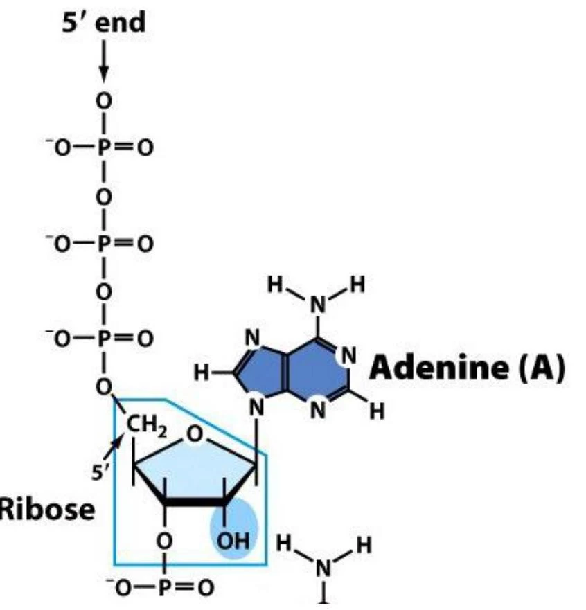
DNA is a nucleic acid composed of repeating units called nucleotides. Each nucleotide is an asymmetric molecule consisting of three distinct components: a deoxyribose sugar, a phosphate group, and a nitrogenous base.
Deoxyribose Sugar: A five-carbon sugar that forms the structural foundation of the nucleotide.
Phosphate Group: Attached to the 5' carbon of the sugar, these groups link nucleotides together.
Nitrogenous Bases: These are the information-carrying components. In DNA, there are four types: Adenine (A), Guanine (G), Cytosine (C), and Thymine (T).
Diagram illustrating the chemical structure of Adenosine Monophosphate (AMP), a nucleotide composed of adenine, ribose sugar, and a phosphate group.
These nucleotides are linked in a long chain through phosphodiester bonds. Specifically, a phosphate group on the 5' carbon of one sugar links to the 3' hydroxyl (-OH) group of the next sugar. This "head-to-tail" connection creates a strand with distinct chemical polarity, characterized by a 5' end and a 3' end.
The Double Helix and Complementary Base Pairing
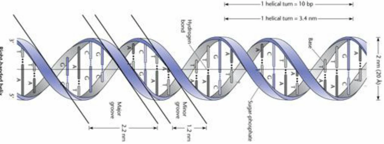
DNA does not exist as a single strand but as a double-stranded molecule. The two strands are held together by hydrogen bonds between the nitrogenous bases of opposing strands. This pairing is highly specific, following the rules of complementary base pairing:
Adenine (A) always pairs with Thymine (T).
Guanine (G) always pairs with Cytosine (C).
Crucially, the two strands are antiparallel, meaning they run in opposite directions. This orientation allows the bases to meet in the center to form the characteristic double helix. The twisting of these strands creates two distinct grooves: a major groove and a minor groove, which are important sites for protein interactions.
Diagram illustrating the structure of a DNA double helix, showing base pairs, sugar-phosphate backbone, major and minor grooves, and dimensions.
DNA Supercoiling and Topoisomerases
In bacteria, the DNA molecule is often a large circle that is much larger than the cell itself. To fit within the cell, the DNA must be highly compacted into a structure called the nucleoid. This is achieved through supercoiling, where the DNA twists upon itself.
However, the process of unwinding DNA for replication or transcription creates intense torsional stress (overwinding). To manage this, cells use enzymes called topoisomerases to modulate supercoiling:
Type I Topoisomerases: These enzymes cut a single strand of the DNA, pass the other strand through the break, and then reseal it, changing the supercoiling state one step at a time.
Type II Topoisomerases: These enzymes cut both strands of the DNA, allowing another segment of the DNA to pass through the break before resealing.
DNA Gyrase: A specific type of Type II topoisomerase found in bacteria that introduces negative supercoils to relieve stress.
The Mechanism of Semi-Conservative Replication
DNA replication is described as semi-conservative. This means that when a double helix is replicated, each of the two resulting daughter molecules consists of one original "old" template strand and one newly synthesized "new" strand.
Replication begins at a specific site known as the origin of replication (oriC), where the DNA "melts" or separates.
Exam Tip: Remember that DNA synthesis always occurs in the 5' → 3' direction. This means new nucleotides can only be added to the 3' hydroxyl (-OH) group of the growing chain.
Key Enzymes in the Replication Fork
As the replication fork opens, a complex assembly of proteins (the replisome) carries out the work:
DNA Helicase (DnaB): Unwinds the double helix at the replication fork.
SSB Proteins (Single-Strand Binding): Bind to the separated strands to prevent them from re-hybridizing (snapping back together).
Primase (DnaG): Synthesizes short RNA primers. Because DNA polymerase cannot start a chain from scratch, it requires the 3'OH group provided by these RNA primers to begin synthesis.
DNA Polymerase III: The primary enzyme responsible for the bulk of DNA synthesis, moving in the 5' → 3' direction.
DNA Polymerase I: Performs two vital roles: it removes the RNA primers using 5' → 3' exonuclease activity and fills the resulting gaps with DNA.
DNA Ligase: The "glue" that seals nicks in the sugar-phosphate backbone by linking 3' hydroxyl groups to adjacent 5' phosphate groups.
Leading vs. Lagging Strands
Because the two strands of the DNA helix are antiparallel and synthesis can only occur in one direction (5' → 3'), the two strands must be replicated differently as the fork moves forward:
The Leading Strand: This strand is synthesized continuously toward the replication fork.
The Lagging Strand: This strand is synthesized discontinuously, moving away from the replication fork. It is produced in short segments called Okazaki fragments. Each fragment requires its own RNA primer to start.
Once the Okazaki fragments are created, RNase H and DNA Polymerase I work together to remove the RNA primers and replace them with DNA. Finally, DNA ligase joins the fragments into a continuous strand.
Summary Table: DNA Polymerase Activities
Activity
Direction
Function
Polymerase
5' → 3'
Synthesizes new DNA strands (Pol I & III)
Exonuclease
5' → 3'
Removes RNA primers (Pol I)
Exonuclease
3' → 5'
Proofreading/error correction (Pol I & III)
Through this highly coordinated enzymatic dance, the cell ensures that its genetic instructions are copied with incredible precision, allowing life to persist across generations.
Keep reading for free.
Create an account to finish this reading and make it part of your study routine.