DNA Repair Mechanisms: Nucleotide Excision and Mismatch Repair
This content explains the essential DNA repair mechanisms, focusing on Nucleotide Excision Repair (NER) for bulky lesions like UV damage and Mismatch Repair (MMR) for correcting replication errors in both prokaryotes and eukaryotes.
The integrity of a genome is constantly under siege. From the moment DNA is replicated to the continuous exposure of the cell to environmental stressors, the double helix is subject to various forms of damage and error. To prevent these issues from becoming permanent mutations that could lead to cell death or uncontrolled growth, organisms have evolved sophisticated DNA repair mechanisms. These pathways are essential for maintaining genomic stability and ensuring that hereditary information is passed accurately from one generation to the next.
Nucleotide Excision Repair and UV Damage
One of the most common threats to DNA is ultraviolet (UV) radiation. UV light can induce specific types of lesions, most notably pyrimidine dimers, where covalent bonds form between adjacent thymine bases on the same DNA strand. These dimers distort the physical structure of the DNA helix, interfering with both replication and transcription.
To combat this, cells utilize nucleotide excision repair (NER). This pathway is designed to recognize and remove bulky lesions that significantly distort the DNA duplex. The process follows a highly coordinated sequence of events:
Recognition: Specific proteins, such as the UVR proteins in bacteria, scan the DNA to identify distortions in the helix.
Excision: Once the damage is identified, an enzyme complex cuts the damaged strand on both sides of the lesion, removing a short segment of single-stranded DNA that includes the error.
Resynthesis: The resulting gap is filled by DNA polymerase, which uses the undamaged complementary strand as a template to restore the original sequence.
Ligation: The final nick in the sugar-phosphate backbone is sealed.
Diagram illustrating the process of nucleotide excision repair in DNA, where UV-damaged DNA is repaired by a complex of proteins including UVR AB, UVR C, and DNA helicase UVR D.
Deficiencies in these repair pathways can have catastrophic consequences. For example, mutations in the UVR A protein can lead to extreme sensitivity to UV light and a significantly increased risk of developing skin cancers.
Even with the high fidelity of DNA polymerase, errors occasionally occur during replication. A base-pair mismatch (such as a G paired with a T) may escape the initial proofreading process. Mismatch Repair (MMR) is the specialized system responsible for identifying and correcting these specific errors.
The fundamental challenge for the MMR system is "strand discrimination." When a mismatch is detected, the repair machinery must determine which of the two strands contains the error (the newly synthesized daughter strand) and which is the correct template (the original parental strand). If the machinery chooses the wrong strand, it will actually "fix" the error by changing the template, thereby cementing the mutation into the genome.
Mismatch Repair in E. coli
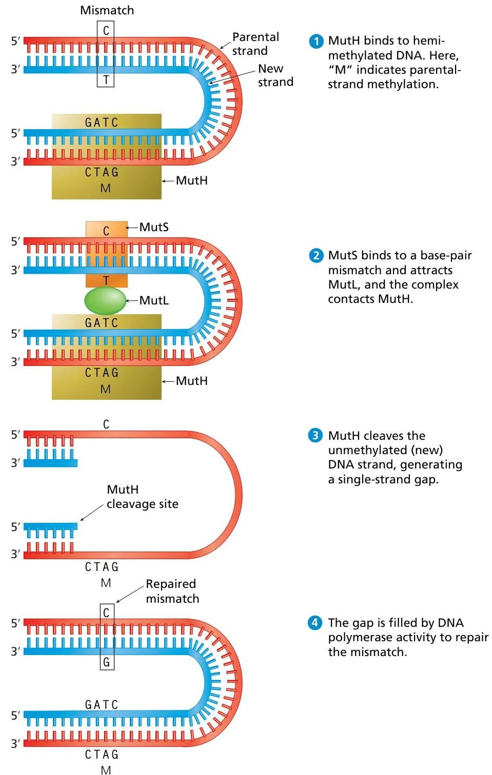
In E. coli, the cell solves the strand discrimination problem through DNA methylation. The parental DNA strands are heavily decorated with methyl groups (CH3) at specific sequences, such as the 5′-GATC-3′ palindrome. Immediately after replication, the newly synthesized daughter strand is not yet methylated. This creates a window of time where the DNA is hemimethylated—the parental strand is methylated, but the daughter strand is not.
The repair process in E. coli relies on three essential proteins:
MutS: This protein acts as a scout, scanning the DNA to recognize and bind directly to the site of the base-pair mismatch.
MutL: Once MutS finds an error, MutL binds to form a complex that coordinates the subsequent repair steps.
MutH: This is a specialized endonuclease. It recognizes the hemimethylated GATC sequence and makes an incision (breaks the phosphodiester bond) specifically on the unmethylated daughter strand.
This diagram illustrates the process of DNA mismatch repair, showing how enzymes like MutH, MutS, and MutL identify and correct errors in DNA replication.
Following the incision by MutH, exonuclease enzymes digest the segment of the daughter strand containing the error. Finally, DNA polymerase fills the gap using the methylated parental strand as a reliable template, ensuring the error is corrected to match the original sequence.
Exam Tip: When studying MMR, always remember the distinction between the parental and daughter strands. In prokaryotes like E. coli, the key to this distinction is the methylation state (hemimethylated DNA).
Mismatch Repair in Eukaryotes
Eukaryotes also possess a highly efficient MMR system, but the mechanism of strand discrimination differs. While eukaryotes utilize proteins that are homologous to MutS and MutL, they do not possess a direct MutH homolog. Instead, they likely use other cues, such as the presence of single-strand breaks (nicks) left behind during lagging strand synthesis, to identify the new strand.
Despite these mechanistic variations, the biological importance of MMR is universal. In humans, defects in eukaryotic MMR genes—such as mutations in $hMLH1$ or $hMLH2$—are directly linked to hereditary nonpolyposis colorectal cancer (HNPCC), also known as Lynch syndrome. This underscores how critical these molecular "spell-checkers" are for preventing the accumulation of mutations that drive oncogenesis.