


Epithelial tissues are essential components of multicellular organisms, serving as the primary interface between the body and its environment. From the skin that protects us from the outside world to the linings of our internal organs, epithelia are specialized sheets of cells that provide structure, protection, and regulation. The evolution of these tissues was a key innovation in Eumetazoa, allowing for compartmentalization—the creation of distinct extracellular compartments like the coelom—and sophisticated physiological regulation, such as specialized digestion and nutrient transport.
The Structure and Polarity of Epithelium
Epithelial cells are not just randomly arranged; they exhibit distinct polarity, meaning they have different functional domains: the apical surface (facing the lumen or exterior), the lateral surface (facing adjacent cells), and the basal surface (resting on the underlying tissue).

To function as an effective barrier, the integrity of the epithelium must be maintained both laterally and basally.
Lateral Integrity: Intercellular Bridges
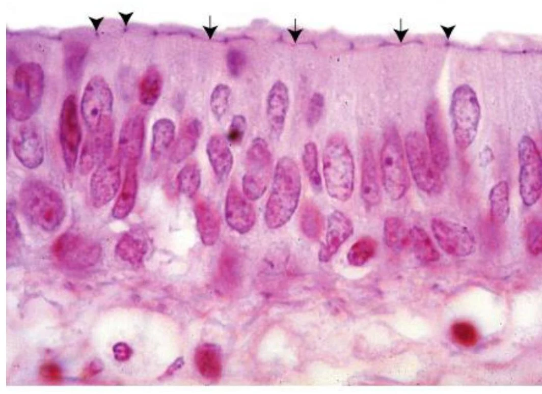
Cells are held together side-to-side by a "junctional complex" known as the terminal bar. These junctions include:
- Tight (Occluding) Junctions (ZO): Composed of proteins like occludin, claudin, and JAM, these create a diffusion barrier that prevents water and compounds from leaking between cells, thereby establishing functional domains in the plasma membrane.
- Anchoring Junctions: These provide mechanical strength.
- Zonula Adherens (ZA): Uses the E-cadherin-catenin complex to attach to the cell's actin filaments.
- Macula Adherens (Desmosomes, MA): Uses cadherins (desmocollin and desmoglein) to attach to intermediate filaments, providing significant mechanical stability and force dissipation.
- Gap Junctions: Formed by connexins, these allow direct communication between cells by permitting the passage of ions and metabolites.
Keep reading for free.
Create an account to finish this reading and make it part of your study routine.
Continue reading freeAlready have an account? Log in
Updated