Distinguishing Structural vs. Functional Foot Deformities
Learn to differentiate between functional and structural foot deformities using the Neutral Calcaneal Stance Position, a key clinical assessment technique for foot and ankle evaluation.
In the clinical assessment of the foot and ankle, one of the most critical challenges is distinguishing between functional movement patterns and true structural deformities. When a patient presents with a collapsed arch or a tilted heel, a clinician must determine if that position is a result of soft tissue laxity or a permanent bony misalignment. To achieve this distinction, we utilize the Neutral Calcaneal Stance Position.
By learning how to manually align the subtalar joint in a non-weight-bearing position, clinicians can identify structural compensations that might otherwise be masked by the patient's weight-bearing mechanics.
Understanding the Subtalar Joint
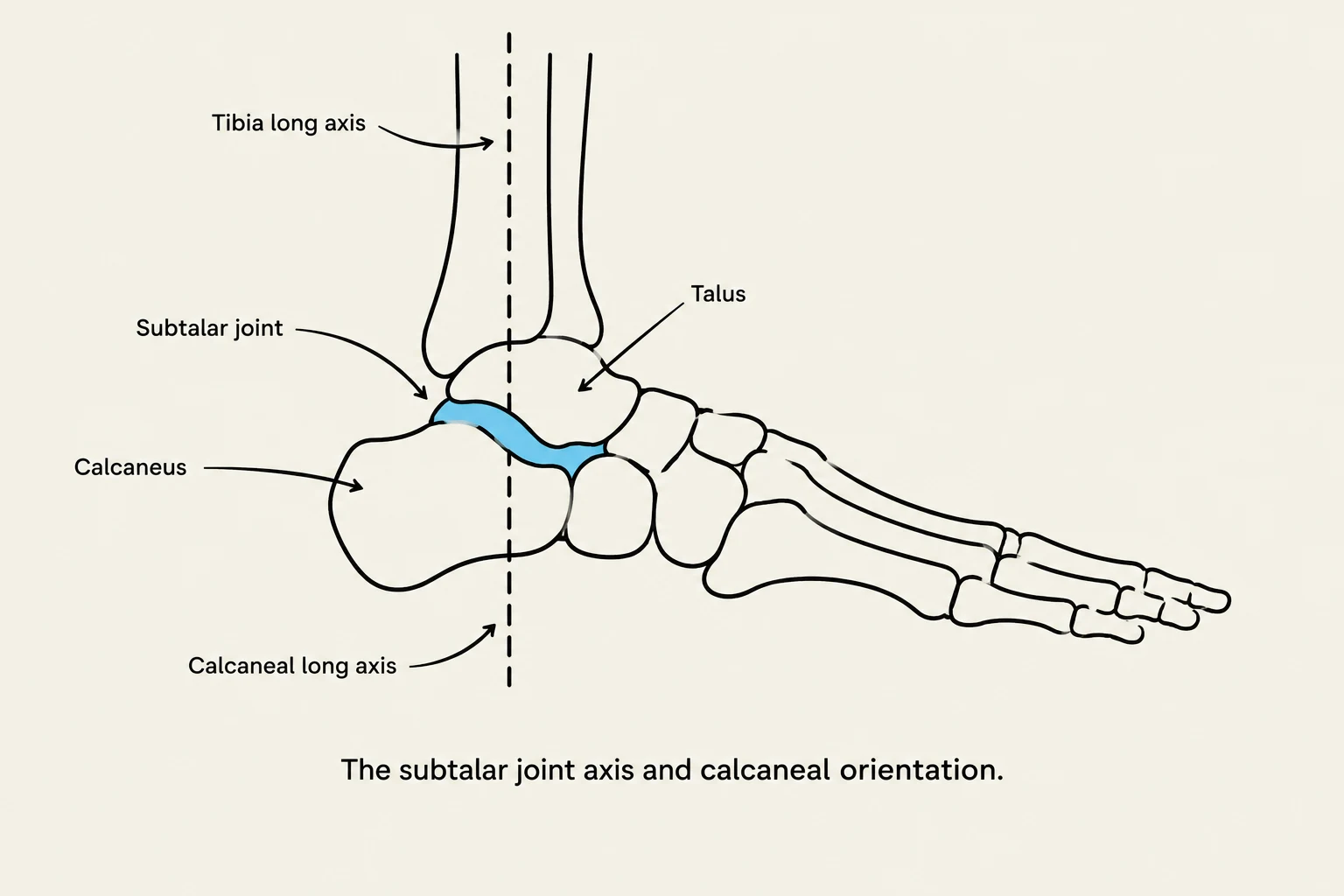
Diagram illustrating the subtalar joint axis and calcaneal orientation.
The subtalar joint is the primary driver of inversion and eversion at the foot. Because the foot is a complex arrangement of bones designed to transition from a mobile adapter to a rigid lever, the position of the calcaneus (the heel bone) serves as the foundation for the entire kinetic chain.
When we assess a patient in a standing position, gravity and muscle tone influence the calcaneal position. A patient might appear to have excessive pronation (valgus) simply because their calf muscles are weak or their midfoot is hypermobile. However, if the calcaneus cannot be brought into a neutral position manually, we are likely looking at a structural issue rather than a functional one.
The Goal of Neutral Alignment
The "Neutral Calcaneal Stance" refers to a specific orientation of the calcaneus where the subtalar joint is neither in excessive eversion (valgus) nor excessive inversion (varus).
In a clinical setting, finding this neutral position involves:
Non-Weight-Bearing Assessment: The patient is typically seated or supine to remove the influence of ground reaction forces and postural compensations.
Manual Manipulation: The clinician uses their hands to palpate the posterior aspect of the calcaneus, guiding it into a position where the axis of the subtalar joint is vertically aligned with the long axis of the lower leg.
Clinical Tip: Always perform your neutral alignment assessment in a non-weight-bearing position first. If you only assess the foot while the patient is standing, you may mistake a functional pronation pattern for a structural deformity.
Identifying Structural vs. Functional Compensations
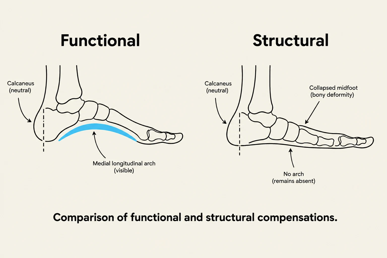
Comparison of functional and structural foot compensations, illustrating the difference between a visible medial longitudinal arch and a collapsed midfoot.
Once you have successfully manipulated the calcaneus into a neutral position, you must observe how the rest of the foot responds. This is where the "identification of structural compensations" occurs.
1. Functional Compensations
If you can move the calcaneus into a neutral position and the medial longitudinal arch "reappears" or the midfoot bones move into a more anatomical alignment, the patient's previous deviation was functional. This suggests that the issue is likely related to muscle weakness, ligamentous laxity, or poor neuromuscular control.
2. Structural Compensations
If, despite your best efforts to align the calcaneus into a neutral position, the midfoot remains collapsed, the toes remain abducted, or the bones of the forefoot remain in a deviated position, you are witnessing a structural compensation. In these cases, the bony architecture itself has adapted to a malalignment, meaning the deformity is "locked" into the skeletal structure.
Clinical Application: A Worked Example
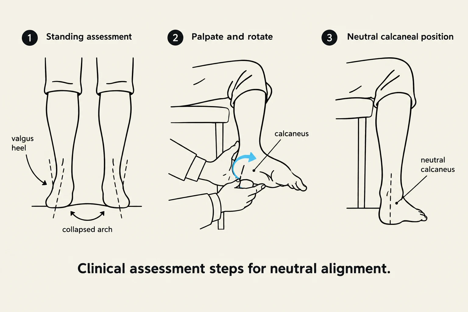
Clinical assessment steps for neutral alignment of the foot, illustrating standing assessment, palpation and rotation, and the neutral calcaneal position.
Imagine a patient presenting with "flat feet" and chronic medial ankle pain.
Step 1: Observe the patient standing. The calcaneus appears in significant valgus (tilted outward), and the arch is completely absent.
Step 2: Move the patient to a seated position. Manually grasp the calcaneus and rotate it into a neutral, vertical position.
Step 3 (The Test):
Scenario A: As you neutralize the heel, the arch rises and the first metatarsal aligns with the midfoot. Conclusion: Functional pes planus (likely due to posterior tibialis weakness).
Scenario B: As you neutralize the heel, the midfoot remains flat and the bones of the tarsals appear crowded or shifted. Conclusion: Structural pes planus (likely due to bony morphology or long-standing ligamentous remodeling).
By distinguishing between these two, the clinician can decide whether the treatment should focus on strengthening and proprioception (functional) or orthotics and surgical consultation (structural).
Summary Table for Assessment
Feature
Functional Deviation
Structural Deviation
Response to Neutralization
Arch rises; bones realign
Arch remains collapsed; bones stay deviated
Primary Cause
Muscle weakness / Laxity
Bony morphology / Remodeling
Clinical Focus
Rehabilitation / Strengthening
Support / Structural correction
Keep reading for free.
Create an account to finish this reading and make it part of your study routine.