


Myeloproliferative Neoplasms (MPNs) represent a group of clonal hematopoietic disorders characterized by the autonomous overproduction of one or more mature blood cell lineages. While these diseases share a common underlying pathophysiology—stemming from mutations in hematopoietic stem cells—they are clinically and morphologically distinct.
To master this topic, one must move beyond simply knowing the names of the diseases. You must be able to differentiate between Polycythemia Vera (PV), Essential Thrombocythemia (ET), and Primary Myelofibrosis (PMF) by analyzing three specific pillars: the dominant cell lineage involved, the clinical presentation (symptoms and risks), and the specific findings in the peripheral blood and bone marrow.
Polycythemia Vera (PV): The Hyperviscous State
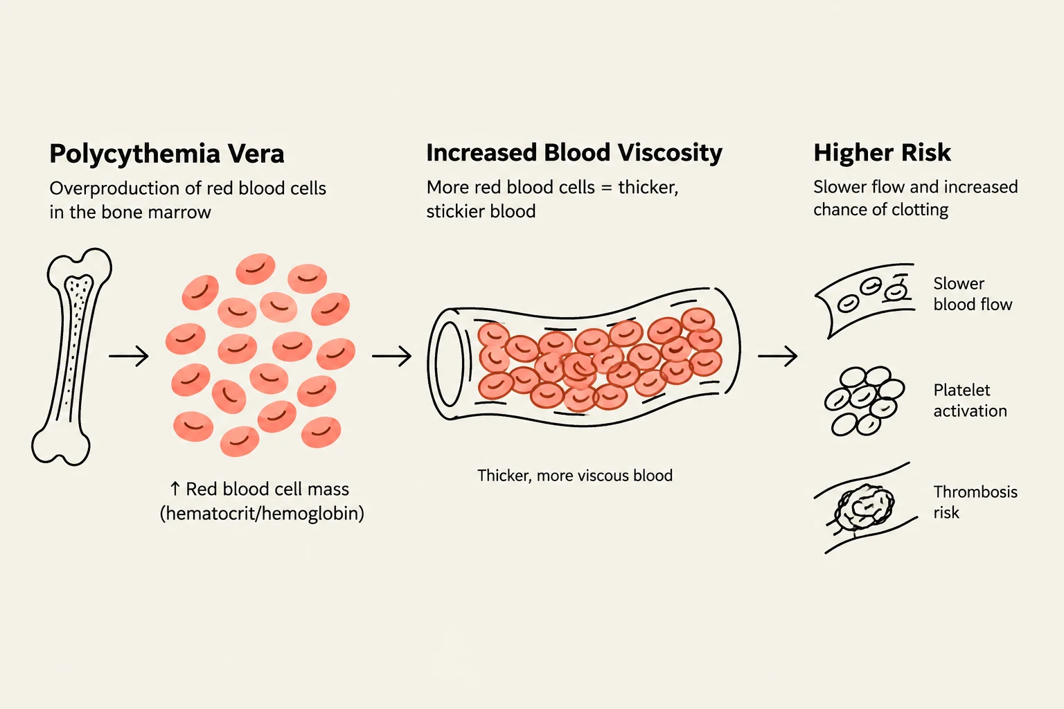
Polycythemia Vera is defined by the excessive production of red blood cells (erythrocytosis), though it often involves increases in white blood cells and platelets as well. The hallmark of PV is a significant increase in hemoglobin and hematocrit levels.
Clinically, the primary concern in PV is hyperviscosity. As the blood becomes "thicker" due to the high red cell mass, the risk of thrombosis (clots) increases significantly. Patients often present with symptoms related to sluggish blood flow, such as headaches, dizziness, or visual disturbances. A classic physical exam finding is aquagenic pruritus—intense itching following a warm bath—which is highly suggestive of PV.
In the bone marrow, you will observe panmyelosis, meaning there is an increase in all three cell lines (erythroid, myeloid, and megakaryocytic).
Keep reading for free.
Create an account to finish this reading and make it part of your study routine.
Continue reading freeAlready have an account? Log in
Updated