Learn to differentiate between oral erosions and ulcers based on their depth of epithelial loss, healing characteristics, and clinical presentation. This guide covers systematic observation, subjective symptoms, and differential diagnosis in oral medicine.
In the clinical practice of oral medicine, the ability to precisely describe and differentiate between various mucosal lesions is a fundamental skill. One of the most common points of confusion for clinicians and students alike is the distinction between an oral erosion and an oral ulcer. While they may appear similar to the untrained eye, the difference in their depth and their healing characteristics provides critical diagnostic clues that can steer a clinician toward a benign diagnosis or a life-threatening malignancy.
Erosion versus Ulcer: The Role of Epithelial Thickness
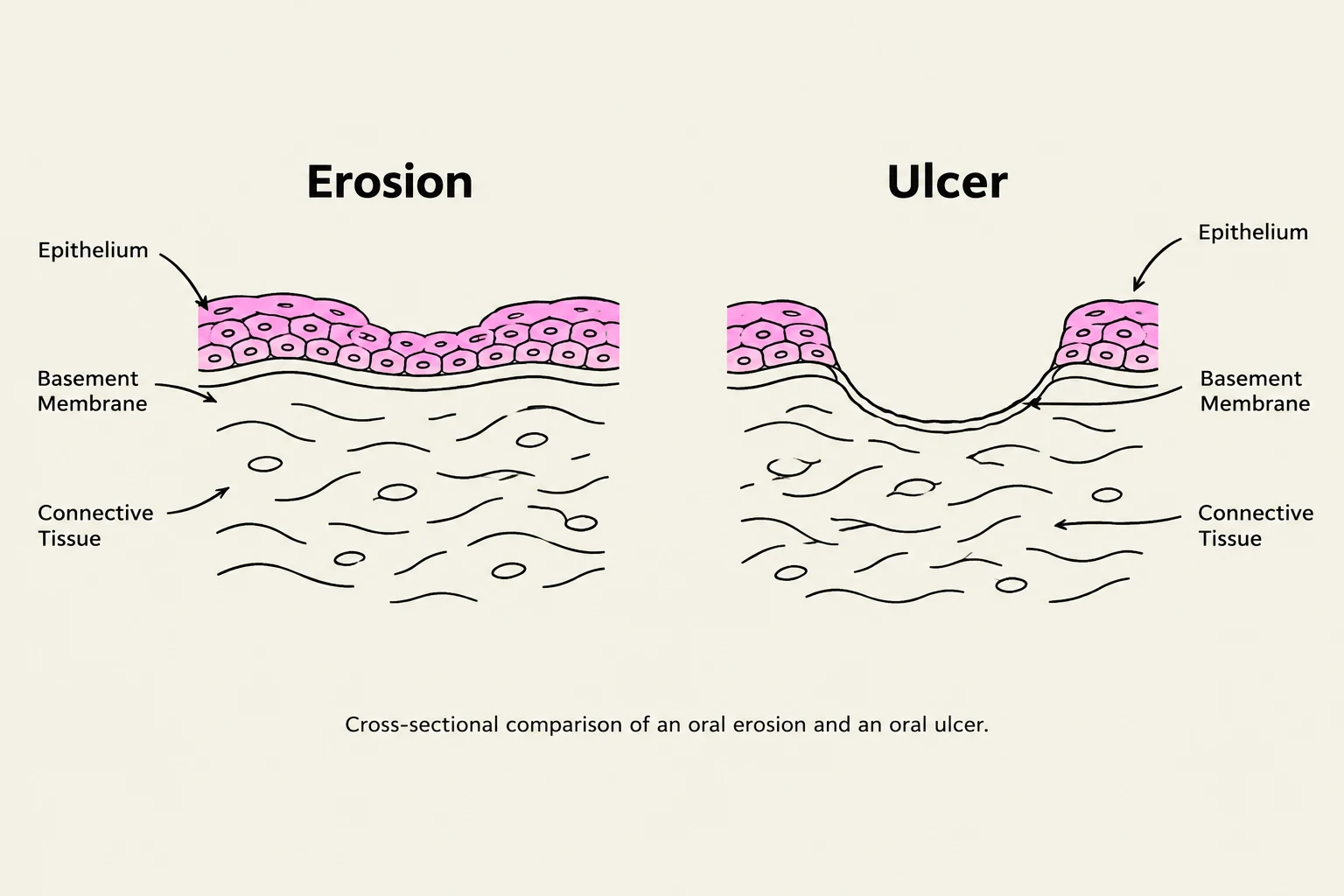
This diagram illustrates the histological differences between an oral erosion and an oral ulcer, showing the layers of epithelium, basement membrane, and connective tissue.
The primary distinction between an erosion and an ulcer lies in the depth of tissue involvement, specifically regarding the loss of the epithelial layer.
Oral Erosions
An oral erosion is characterized by a partial loss of epithelium. These lesions often occur as a secondary change following the rupture of a primary lesion, such as a vesicle (small fluid-filled sac) or a bulla (a larger fluid-filled sac). Because the damage is superficial and does not penetrate through the full thickness of the epithelium into the underlying connective tissue, erosions typically heal without leaving a scar.
Oral Ulcers
In contrast, an oral ulcer involves a full loss of thickness in the epithelium. An ulcer penetrates through the entire epithelial layer, often reaching the basement membrane or the underlying connective tissue. Because the lesion reaches these deeper structures, an ulcer may leave a scar upon healing if the depth of the lesion is significant.
Clinical Pearl: If a patient describes a "blister" that has since popped, you are likely observing a secondary erosion.
Systematic Clinical Observation
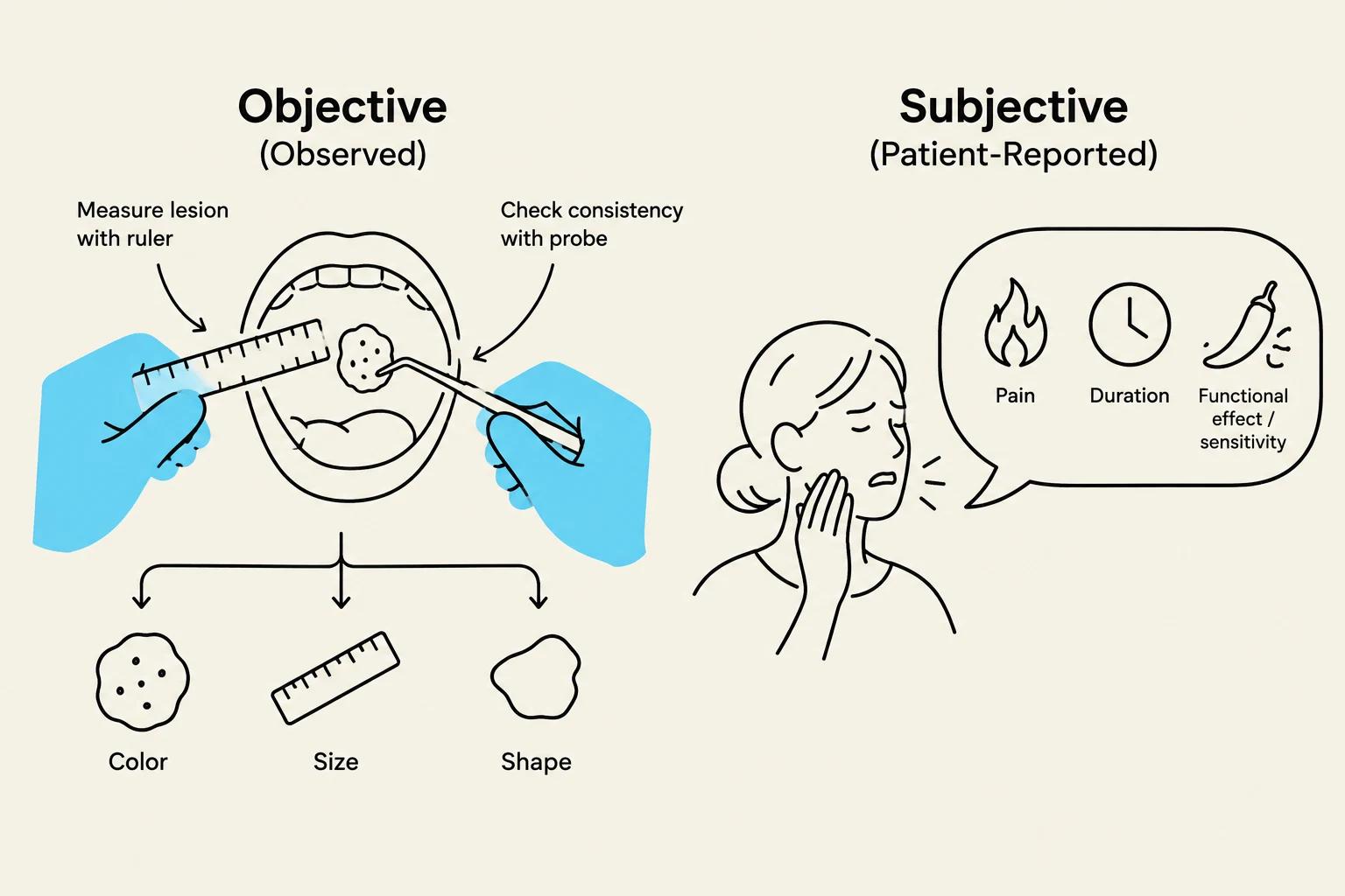
This illustration depicts the objective and subjective assessment of a lesion, likely in a medical context. Objective assessment involves measuring the lesion's size, color, and shape, and checking its consistency. Subjective assessment, reported by the patient, includes pain, duration, and functional effects or sensitivity.
To move from a simple observation to a professional clinical assessment, a clinician must separate observed findings (objective signs) from patient-reported symptoms (subjective sensations).
Objective Observed Findings
When documenting a lesion, you must be as precise as possible using standardized morphological language. A complete description should include:
Site and Dimensions: Record the exact anatomical sub-site (e.g., left buccal mucosa) and measure the longest dimension and perpendicular width, ideally in millimeters.
Number and Distribution: Is the lesion solitary, multiple, bilateral, or diffuse (scattered like sand)? Note that bilateral lesions are often less likely to be neoplastic (cancerous) than solitary ones.
Color and Surface: Describe the color (white, red, pigmented, or "variegated"/speckled) and the surface texture (smooth, rough, reticular/lacy, or crusted).
Borders and Base: Note if the margins are well-defined or poorly defined, and whether the base is flat or pedunculated (on a stalk).
Consistency: Palpate the tissue to determine if it is soft, fluctuant, firm, or indurated.
Subjective Patient-Reported Symptoms
The patient's history provides the "sequence" of the disease. You must ask:
Pain and Sensation: Is there burning or sharp pain? (Note: While both herpes and aphthous ulcers are painful, clinical experience often suggests herpes may present with higher pain levels, though location is a more reliable differentiator).
Duration and Recurrence: When did it start? Does it come back in the same spot or different places? This helps identify patterns like Recurrent Aphthous Stomatitis (RAS).
Functional Effects: Can the patient eat normally, or must they avoid hot, spicy, or acidic foods?.
Differential Diagnosis and Clinical Reasoning
Illustration showing a lateral border lesion on the tongue and a magnified view of firm/hard tissue compared to surrounding soft tissue, with a gloved finger palpating for induration.
A structured diagnostic process requires a "ranked differential." This means listing at least three possible diagnoses, starting with the most likely, and providing a specific reason for each.
Example Case: The Suspicious Tongue Lesion
Imagine a patient presents with a solitary, irregular, red-and-white lesion on the lateral border of the tongue that feels hard to the touch.
Squamous Cell Carcinoma: Ranked first due to the patient's age, smoking history, and the presence of induration (firmness/hardness), which is a major red flag for malignancy.
Traumatic Ulcer: A possibility if there is a sharp tooth or denture flange rubbing the area, though the induration makes this less likely.
Erythroleukoplakia: A descriptive term for a red and white lesion where the exact pathology is not yet known.
Exam Tip: The "Magic Number"
If you identify a potential traumatic cause (like a sharp tooth) and treat it (by smoothing the tooth), you must follow up with the patient in two weeks to see if the lesion has healed.
Investigation and Follow-up
If a lesion is severe, atypical, or persistent, you must propose targeted investigations.
The Importance of Biopsy
A biopsy is often required for a definitive diagnosis. However, clinicians must be wary of negative adjunct tests. If a lesion looks clinically suspicious (e.g., the patient is losing weight, has induration, or has a history of smoking) but the biopsy comes back negative, you should not ignore your clinical sense. A negative result might simply mean the biopsy sample was taken from an area of the lesion that was not representative of the pathology. In such cases, you must reassess and potentially perform a repeat biopsy of a different area.
Follow-up Protocol
Follow-up is an integral part of the diagnostic process. Whether you are monitoring a suspected traumatic ulcer or waiting for a specialist referral, you must ensure the patient returns to confirm healing or to receive definitive results.
Feature
Oral Erosion
Oral Ulcer
Epithelial Loss
Partial (Superficial)
Full thickness
Connective Tissue
Not exposed
Exposed
Scarring
Heals without scar
May leave a scar
Keep reading for free.
Create an account to finish this reading and make it part of your study routine.