


The human visual system is a sophisticated network that converts light energy into neuronal signals through the retina and processes them via complex pathways to the brain. While much of clinical ophthalmology focuses on the integrity of the retina and optic nerve, a significant portion of visual dysfunction arises from disorders of the ocular motor system. These disorders can manifest as drooping eyelids, double vision, or involuntary eye movements, often serving as critical windows into the health of the cranial nerves and brainstem.
Blepharoptosis
Blepharoptosis, or ptosis, refers to the abnormal drooping of the upper eyelid. Because the eyelid position is maintained by both muscle and nerve, clinicians must categorize ptosis based on its underlying mechanism to narrow the differential diagnosis.
Classification of Ptosis
Ptosis is broadly classified into four categories:
- Mechanical Ptosis: Caused by physical factors such as skin redundancy or the presence of a mass.
- Aponeurotic Ptosis: Resulting from the stretching or dehiscence of the levator aponeurosis.
- Myogenic Ptosis: Associated with primary muscle disorders, such as myasthenia gravis or oculopharyngeal muscular dystrophy.
- Neurogenic Ptosis: Caused by lesions affecting the levator palpebrae superioris or Müller's muscle.
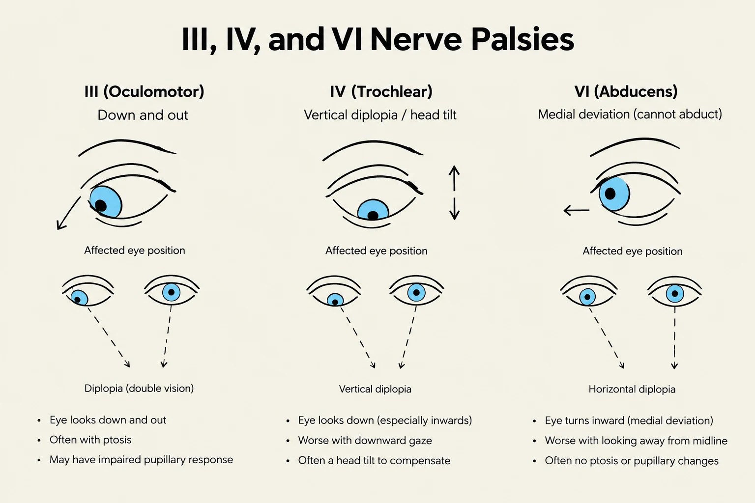
Exam Tip: The Pupil Clue When evaluating neurogenic ptosis, always examine the pupils. A smaller pupil (miosis) suggests Horner's syndrome (sympathetic paresis), whereas a larger or normal-sized pupil suggests an oculomotor nerve (III) palsy (parasympathetic deficit).
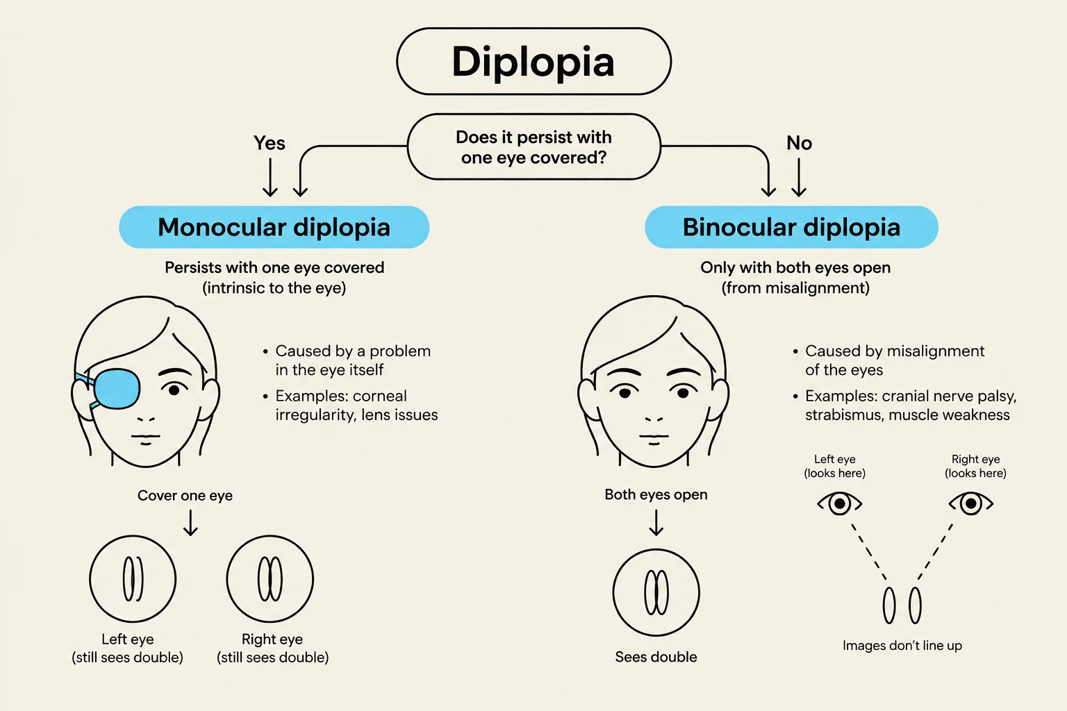
Diplopia: Monocular vs. Binocular
Diplopia, or double vision, is one of the most common complaints in ocular motility disorders. The first step in clinical assessment is determining whether the diplopia is monocular or binocular.

Keep reading for free.
Create an account to finish this reading and make it part of your study routine.
Continue reading freeAlready have an account? Log in
Updated