


Effective neural communication relies on the precise movement of ions and the timely release of chemical messengers across synapses. When this delicate balance is disrupted—whether by an autoimmune attack on receptors or a genetic defect in an ion channel—the result is a spectrum of neurological and systemic diseases. These disorders are broadly categorized into pathologies affecting the neuromuscular junction (NMJ) and channelopathies, which involve the dysfunction of ion channels themselves.
The Fundamentals of Synaptic Transmission
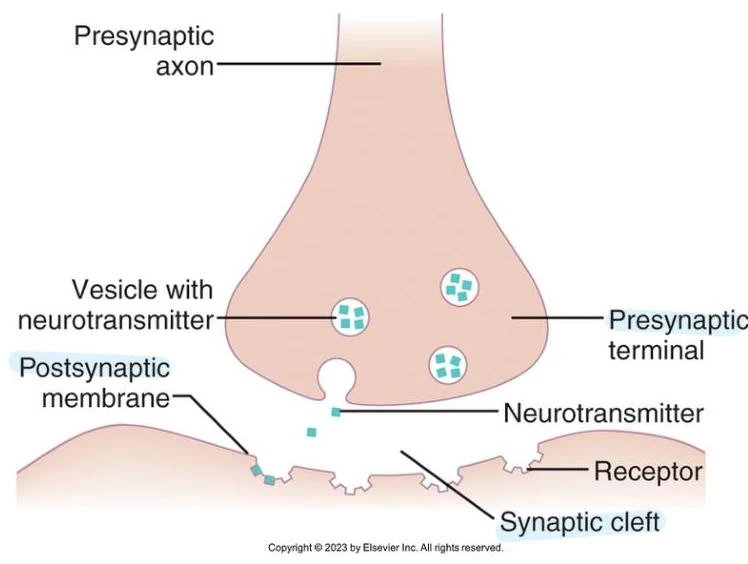
To understand these disorders, one must first understand the healthy functioning of a synapse. A chemical synapse consists of three primary components: the presynaptic terminal (the sending neuron), the synaptic cleft (the gap between cells), and the postsynaptic membrane (the receiving cell).
The process of neurotransmission follows a specific sequence:
- An action potential reaches the presynaptic axon terminal.
- This electrical change triggers the opening of voltage-gated channels, allowing calcium to flow into the cell.
- The influx of promotes the movement of synaptic vesicles toward the membrane, where they release neurotransmitters into the synaptic cleft.
- The neurotransmitter diffuses across the cleft and binds to specific receptors on the postsynaptic membrane.
Depending on the neurotransmitter and the receptor type, this binding results in a change in the postsynaptic membrane potential. An Excitatory Postsynaptic Potential (EPSP) causes depolarization (making the cell more positive), while an Inhibitory Postsynaptic Potential (IPSP) causes hyperpolarization (making the cell more negative).
At the neuromuscular junction (NMJ), which connects the peripheral nervous system (PNS) to skeletal muscle, the neurotransmitter acetylcholine (ACh) is released. ACh binds to nicotinic receptors on the muscle cell, triggering the rapid depolarization necessary for muscle contraction.
Keep reading for free.
Create an account to finish this reading and make it part of your study routine.
Continue reading freeAlready have an account? Log in
Updated