An overview of the renal and urinary systems, including normal anatomy and physiology, common diseases, and urinary incontinence. Covers conditions like kidney failure, infections, stones, and different types of incontinence.
The renal and urinary systems are vital for maintaining homeostasis, regulating blood pressure, and filtering metabolic waste from the body. Because these systems are highly interconnected, a pathology in one structure often cascades to affect others. Understanding the transition from normal physiology to clinical disease is essential for diagnosing conditions ranging from simple infections to end-stage renal disease (ESRD).
Normal Renal Anatomy and Physiology
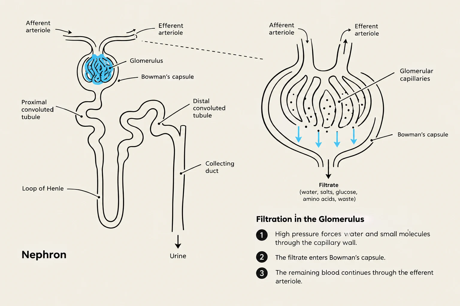
The kidneys serve as the primary filtration units of the body. To understand disease, one must first understand the functional unit of the kidney: the nephron. Within the nephron lies the glomerulus, a specialized network of capillaries designed for ultrafiltration. The glomerulus acts as a biological sieve, allowing water and small solutes to pass through while retaining larger proteins and blood cells within the vascular system.
Diagram illustrating the structure of a nephron and the process of filtration in the glomerulus.
The urinary tract also includes the structures responsible for transporting and storing urine, such as the ureters, bladder, and urethra. The regulation of this system involves a complex interplay between the autonomic nervous system and the muscular components of the bladder.
The Autonomic Control of Micturition
The process of storing and emptying urine is governed by two primary branches of the autonomic nervous system:
Parasympathetic Nervous System (PNS): Facilitates micturition (voiding). During this phase, the detrusor muscle (the bladder wall) contracts, and the internal sphincter relaxes.
Sympathetic Nervous System (SNS): Facilitates urine storage. During this phase, the detrusor muscle relaxes, and the internal sphincter remains tense.
Somatic Nervous System: Provides voluntary control over the external sphincter.
The Language of Renal Disease
Clinicians use specific terminology to describe abnormalities found during urinalysis or patient interviews. Recognizing these terms is the first step in identifying renal pathology.
Dysuria: Painful or difficult urination.
Pyuria: The presence of pus (white blood cells) in the urine, often indicating infection.
Hematuria: The presence of blood in the urine. This can be "occult" (hidden and only detectable via testing) or visible.
Nocturia: The need to wake up during the night to urinate.
Proteinuria: The presence of excess proteins in the urine, which often suggests damage to the glomerular filter.
Glycosuria: The presence of glucose in the urine, frequently associated with diabetes mellitus.
Azotemia: A clinical syndrome characterized by renal failure with abnormal blood tests, specifically elevated Blood Urea Nitrogen (BUN) levels.
Major Categories of Urinary Disease
Diseases of the urinary system can be categorized by their origin and their impact on renal function.
Renal Failure and Glomerular Disease
Renal failure is often classified by its onset and duration. Acute renal failure involves a rapid decline in function, often marked by sudden changes in BUN levels. In contrast, chronic renal failure is a long-term decline that can progress to End-Stage Renal Disease (ESRD), requiring interventions such as hemodialysis.
Common drivers of chronic disease include:
Chronic glomerulonephritis: Inflammation of the glomeruli.
Diabetes Mellitus (DM): A leading cause of kidney damage.
Hypertension (HTN): High blood pressure is a common cause of kidney disease.
Many primary glomerular diseases are triggered by autoimmune reactions. Additionally, diseases of the renal vasculature (the blood vessels supplying the kidney) can significantly impair function.
Obstruction, Infection, and Stones
The urinary tract is particularly susceptible to bacterial urinary tract infections (UTI). Other mechanical issues include:
Nephrolithiasis: The formation of kidney stones.
Urinary tract obstruction: Any blockage in the flow of urine, which can lead to pressure buildup and kidney damage.
Exam Tip: When evaluating a patient with renal issues, always check for "major determinants." Remember that high blood pressure and autoimmune reactions are primary drivers of glomerular disease.
Urinary Incontinence
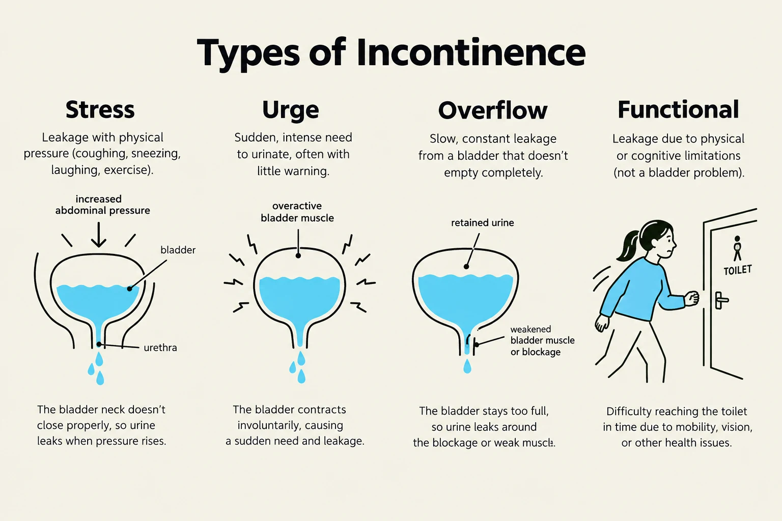
Urinary incontinence is the involuntary loss of urine. It is categorized based on the underlying mechanism of the failure to control the bladder or sphincter.
This diagram illustrates four types of incontinence: stress, urge, overflow, and functional, along with their causes and symptoms.
Types of Incontinence
Stress Incontinence: Leakage occurs during physical exertion that increases intra-abdominal pressure, such as laughing, coughing, sneezing, or lifting. This is often due to weakness in the pelvic floor muscles.
Urge Incontinence: Characterized by a sudden, intense need to urinate followed by involuntary loss of urine. This is often linked to "overactive bladder syndrome" or inappropriate signals from the Central Nervous System (CNS).
Overflow Incontinence: Occurs when the bladder cannot empty completely, causing it to "overflow".
Functional Incontinence: Occurs when physical or cognitive impairments prevent a person from reaching the toilet in time, even if the urinary system itself is functioning.
Neurogenic Bladder: A condition where neurological damage (to the spinal cord or brain) disrupts the signals between the brain and the bladder.
Management and Treatment
Treatment strategies vary depending on the type of incontinence and may include:
Lifestyle modifications: Managing water and caffeine intake.
Bladder retraining: Establishing a regular voiding schedule.
Physical therapy: Including Kegel exercises (pelvic floor strengthening), biofeedback, or electrical stimulation.
Surgical intervention: For more severe cases.
Clinical Case Study: Rehabilitation Perspective
In a clinical setting, such as rehabilitation, understanding the patient's renal status is critical for safety and care planning.
Case: A 74-year-old male presents with ESRD and is currently undergoing hemodialysis. He has a right upper extremity (RUE) shunt/fistula for dialysis access. He is currently in rehab, ambulating 10 feet with a front-wheeled walker (FWW) and moderate assistance. He shows muscle strength (MMT) ranging from 3/5 to 4/5 in his bilateral lower extremities and has poor tolerance to activity.
Clinical Priorities:
When treating this individual, the therapist must consider:
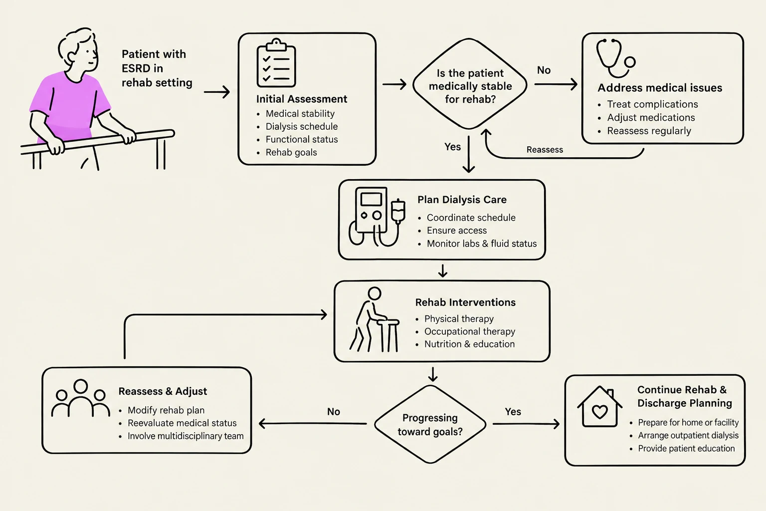
Dialysis Schedule: The patient's energy levels and metabolic state will fluctuate based on his MWF (Monday, Wednesday, Friday) dialysis schedule.
Vascular Access: Protecting the RUE shunt/fistula is paramount; avoid blood pressure cuffs or heavy lifting on that arm.
Activity Tolerance: Due to ESRD, the patient may experience fatigue or electrolyte imbalances that affect strength and endurance.
Flowchart illustrating the rehabilitation process for patients with End-Stage Renal Disease (ESRD), including initial assessment, dialysis care planning, interventions, reassessment, and discharge planning.
Keep reading for free.
Create an account to finish this reading and make it part of your study routine.