Digestive System Microanatomy and Comparative Anatomy
An overview of the microanatomy and comparative anatomy of the digestive system in veterinary medicine, covering tissue organization, embryonic origins, and variations across species.
The gastrointestinal (GI) system is a complex, highly organized series of organs designed to transform food into usable energy and building blocks for the body. From the moment of ingestion to the final excretion of waste, the system utilizes a combination of mechanical forces, enzymatic reactions, and specialized cellular structures to maintain homeostasis. Understanding the microanatomy of these organs—specifically how their tissue layers are organized and how they vary across different species—is fundamental to veterinary medicine.
Functions of the Gastrointestinal System
The GI tract is not merely a tube for transport; it is a multifunctional biological factory. Its primary roles include:
Prehension: The process of ingestion and the initial fragmentation of food.
Digestion: The mechanical and enzymatic breakdown (or fermentation in some species) of nutrients.
Absorption: The movement of nutrients from the lumen into the bloodstream or lymphatic system.
Synthesis and Secretion: The production of digestive enzymes, mucus, and various hormones.
Excretion: The removal of undigested waste material from the body.
Embryological Origins of Digestive Tissues
The diverse tissues found within the digestive tract arise from different embryonic germ layers. This developmental origin dictates the specific function and structure of each layer:
Endoderm: Gives rise to the epithelium (mucosa) that lines the lumen, as well as the parenchymatous organs such as the liver and pancreas and their associated glands.
Mesoderm: Forms the structural and contractile components, including the submucosa, the muscle layers (muscularis), the serosa, and the mesentery.
Ectoderm: Contributes to the stratified squamous epithelium found in the oral cavity and anus, as well as the nerve plexi that regulate motility.
General Organization of Tubular Digestive Organs
Most digestive organs follow a consistent "four-tunic" structural pattern. This organization allows the organ to protect itself, secrete necessary substances, and move food efficiently.
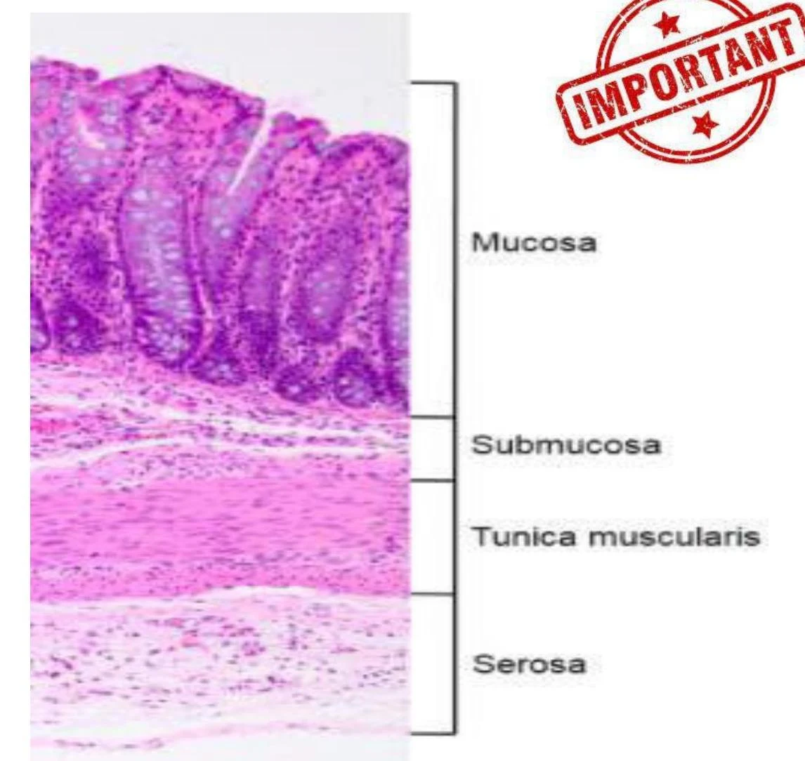
1. Mucosa
The innermost layer, in direct contact with food, consists of three sub-layers:
Epithelium: The functional lining (e.g., simple columnar for absorption or stratified squamous for protection).
Lamina Propria: Loose connective tissue containing blood vessels, lymphatics, and immune cells that support the epithelium.
Muscularis Mucosae: A thin layer of smooth muscle that facilitates local movement of the mucosa.
2. Submucosa
A layer of loose connective tissue that facilitates the motility of the mucosa. It houses larger blood vessels, lymphatic nodules, and the submucosal nerve plexus. In some areas, such as the esophagus, it may contain submucosal glands.
3. Muscularis (Muscularis Externa)
Typically composed of two layers of smooth muscle: an inner circular layer and an outer longitudinal layer. This arrangement controls the size of the lumen and the peristaltic contractions that move food along the tract. The myenteric nerve plexus is located between these two layers to coordinate movement.
4. Serosa and Adventitia
The outermost covering depends on the organ's location:
Serosa: Consists of a mesothelium and loose connective tissue. It is found on organs within the peritoneal, pleural, or pericardial cavities (e.g., the peritoneum).
Adventitia: Consists of only connective tissue. It is found on organs that do not border these cavities, such as the cervical esophagus, where it blends with surrounding fascia.
Histological cross-section of the intestinal wall showing the mucosa, submucosa, tunica muscularis, and serosa layers.
Lining Epithelium Types
The type of epithelium present in a specific part of the GI tract is a direct reflection of its physiological role:
Stratified Squamous Epithelium: This multi-layered tissue is designed for protection against abrasion. It is found in the oral cavity, the esophagus, and the anus. It can be keratinized (found in species consuming coarse food, like horses or ruminants) or non-keratinized (found in carnivores like dogs and cats).
Simple Columnar Epithelium: This single layer of tall cells is optimized for secretion and absorption. It lines the glandular stomach and the intestines.
Comparative Anatomy of the Oral Cavity and Dentition
The oral cavity, including the lips, cheeks, palate, pharynx, and tongue, serves as the site for prehension and initial mechanical breakdown.
The Tongue and Papillae
The tongue is covered by stratified squamous epithelium, which is typically keratinized on the dorsal surface to withstand the friction of food. The surface is irregular due to various papillae:
Filiform papillae: Slender and conical; primarily provide mechanical function.
Circumvallate papillae: Large, flat, and surrounded by a sulcus; contain taste buds.
Foliate papillae: Folds of mucosa located near the root of the tongue; contain taste buds.
Dentition and Tooth Structure
Teeth are specialized for the specific diet of the animal. A tooth consists of hard tissues (enamel, dentin, and cementum) and soft tissues (pulp and periodontal ligament). The periodontium—comprising the gingivae, periodontal ligament, cementum, and alveolar bone—works to resist the forces applied during mastication.
Morphologically, teeth are categorized into two types:
Brachydont: Short teeth that cease to grow after eruption (e.g., carnivore teeth, ruminant incisors).
Hypsodont: Tall teeth that continue to grow after eruption (e.g., horse teeth, ruminant cheek teeth).
Salivary Glands and Secretion
Saliva provides moisture, lubrication, and contains enzymes like amylase and lipase to begin digestion. Salivary glands are classified by their product:
Serous: Watery secretion containing enzymes (e.g., the parotid gland).
Mucous: Viscous, slick secretion.
Mixed: Contains both serous and mucous cells, often forming a "serous demilune" (a crescent of serous cells around a mucous acinus).
Glandular structures are organized into tubular (straight/coiled), acinar (pie-shaped), or alveolar (large lumen) adenomeres.
Comparative Gastric Anatomy
The structure of the stomach varies significantly among domestic species, categorized into three main types:
Simple Stomach: Contains only glandular mucosa (e.g., carnivores and humans).
Composite Stomach: Contains both glandular and stratified squamous mucosa (e.g., pigs and horses).
Compound Multichambered Stomach: Characteristic of ruminants, featuring forestomachs and a true stomach.
The Ruminant Stomach
Ruminants possess a complex four-compartment system designed for microbial fermentation:
Rumen: The largest compartment; acts as a fermentation vat where microbes break down cellulose and the lining absorbs volatile fatty acids (VFAs).
Reticulum: Features a "honeycomb" mucosa; it helps in the formation of cuds and acts as a sieve.
Omasum: Known as the "butcher's bible" due to its many leaves; it removes excess water and reduces particle size.
Abomasum: The "true stomach"; it has a glandular lining and uses acid and enzymes for digestion.
Exam Tip: When identifying histological sections, remember that the presence of a muscularis mucosae and a submucosa is a hallmark of the tubular GI structure, but the specific type of epithelium (stratified squamous vs. simple columnar) will tell you exactly which part of the tract you are looking at.
Keep reading for free.
Create an account to finish this reading and make it part of your study routine.