An overview of the digestive system's anatomy and physiology, detailing its six functions, the structure of the alimentary canal, the journey of food from mouth to stomach, the roles of accessory organs like the liver, gallbladder, and pancreas, and the final processes of absorption and elimination.
The digestive system functions much like a biological disassembly line. Its primary mission is to process food, extract essential nutrients, and eliminate any non-digestible residue. Because most nutrients in the food we eat are too large to be used by our tissues in their existing form, the system must break down complex polymers into smaller, usable monomers through a series of coordinated mechanical and chemical processes.
The study of the digestive tract and the treatment of its disorders is known as gastroenterology. To understand how this system works, we must look at its two main components: the gastrointestinal (GI) tract (the continuous 30-foot muscular tube from mouth to anus) and the accessory organs that assist in the breakdown process.
The Six Functions of Digestion
The digestive process is not a single event but a sequence of six distinct functional stages:
Ingestion: The intake of food into the mouth.
Secretion: The release of saliva, mucus, digestive enzymes, hormones, and pH buffers.
Mixing & Movement: Moving ingested materials through the tract and mixing them with secretions.
Digestion: The mechanical and chemical breakdown of food.
Absorption: The uptake of nutrient molecules into the cells of the digestive tract and subsequently into the blood.
Excretion: The removal of non-digestible waste.
Study Tip: Mechanical vs. Chemical Digestion
Remember that mechanical digestion (like chewing or churning) physically breaks food into smaller particles to increase surface area, while chemical digestion uses enzymes to perform hydrolysis reactions that break macromolecules into monomers (e.g., turning polysaccharides into monosaccharides).
Anatomy of the Alimentary Canal
The four distinct layers of the alimentary canal wall.
The alimentary canal is a muscular tube composed of four distinct layers, organized from the innermost (deep) to the outermost (superficial):
Mucosa: The innermost lining. It consists of an epithelium (mostly simple columnar, though stratified squamous in the mouth and esophagus), a lamina propria of loose connective tissue, and a muscularis mucosae that creates ridges to increase surface area.
Submucosa: A thick layer of loose connective tissue containing the submucosal plexus, a network of nervous tissue that regulates secretions.
Muscularis externa: Composed of smooth muscle, this layer propels food through the canal via motility. It contains the myenteric plexus.
Serosa: The outermost layer of loose connective tissue that helps anchor the organs.
The combination of the submucosal and myenteric plexuses forms the enteric nervous system, which provides local control of the digestive tract.
The Journey of Food: From Mouth to Stomach
The Mouth and Esophagus
Digestion begins in the oral cavity through mastication (chewing) and the action of saliva. Saliva contains enzymes such as salivary amylase for carbohydrate digestion and lingual lipase for fats. Once food is chewed and moistened, it becomes a bolus.
The bolus travels through the pharynx and into the esophagus, a muscular tube that uses peristalsis—waves of muscular contractions—to move food toward the stomach. Two sphincters act as gates: the upper esophageal sphincter and the lower esophageal sphincter. If the lower sphincter fails to close properly, stomach contents can regurgitate, causing heartburn.
The Stomach
Microscopic view of gastric pits and specialized secretory cells.
The stomach is a muscular sac located in the upper left abdominal cavity. Here, the bolus is transformed into a soupy, semi-digested mixture called chyme. The stomach is highly acidic, maintaining a pH between 1.5 and 3.5.
The stomach wall contains gastric pits which house specialized cells:
Mucus cells: Secrete protective mucus.
Chief cells: Secrete pepsinogen, an inactive enzyme (zymogen) that is converted into pepsin by stomach acid to digest proteins.
Parietal cells: Secrete hydrochloric acid (HCl) to activate enzymes and destroy pathogens, and intrinsic factor, which is essential for absorbing Vitamin B12.
Enteroendocrine cells: Such as G cells, which secrete gastrin to stimulate HCl release.
Accessory Organs: The Chemical Powerhouses
As chyme enters the small intestine, it is met by critical secretions from the liver, gallbladder, and pancreas.
The Liver and Gallbladder
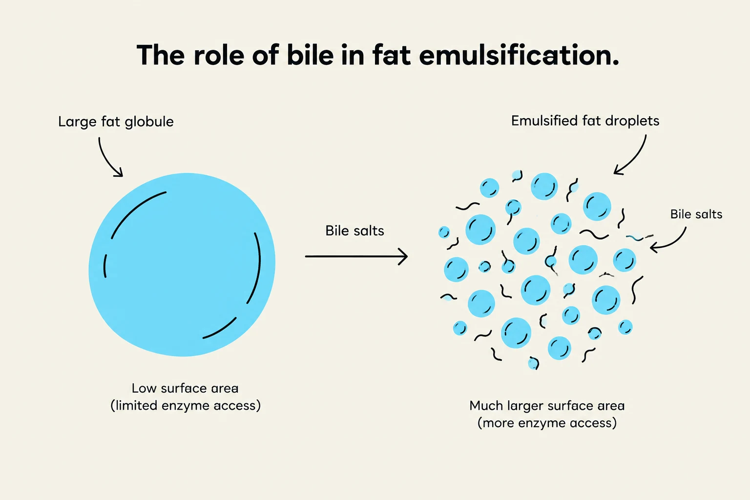
This diagram illustrates how bile salts emulsify large fat globules into smaller droplets, increasing the surface area for enzyme access and digestion.
The liver is the body's largest gland. Its primary digestive role is the production of bile via specialized cells called hepatocytes. Beyond digestion, the liver manages metabolic homeostasis by absorbing glucose, amino acids, and iron, and regulating blood sugar by converting stored glycogen back into glucose.
The gallbladder is a pear-shaped sac that stores and concentrates the bile produced by the liver. Bile contains bile salts (steroids synthesized from cholesterol) which emulsify fats, making them easier for enzymes to digest, as well as bilirubin, a byproduct of hemoglobin decomposition.
The Pancreas
The pancreas is a dual-function gland located posterior to the stomach. While its endocrine portion secretes hormones like insulin and glucagon, its exocrine portion (comprising 99% of the gland) produces pancreatic juice.
Released into the small intestine via the pancreatic duct, this juice contains:
Bicarbonate: A buffer that neutralizes the highly acidic chyme from the stomach.
Trypsinogen: A zymogen converted into trypsin for protein digestion.
Pancreatic amylase: For starch digestion.
Pancreatic lipase: For fat digestion.
Ribonuclease & deoxyribonuclease: For the digestion of RNA and DNA.
Conclusion: Absorption and Elimination
The process concludes in the large intestine, where the remaining material has been stripped of most nutrients. The large intestine focuses on absorbing water and electrolytes, ultimately forming feces for defecation. Efficient nutrient uptake is made possible by the specialized structure of the small intestine, where villi and microvilli maximize the available surface area for absorption.
Keep reading for free.
Create an account to finish this reading and make it part of your study routine.