


When a child presents with a skin eruption, the clinician is often faced with a daunting array of possibilities. Because many pediatric rashes share similar morphological features—such as erythema, papules, or vesicles—the ability to differentiate between them is a core competency in pediatric dermatology. Mastering this process requires more than just memorizing a list of diseases; it requires a systematic approach to morphology, distribution, and clinical context.
The Systematic Approach to Morphology
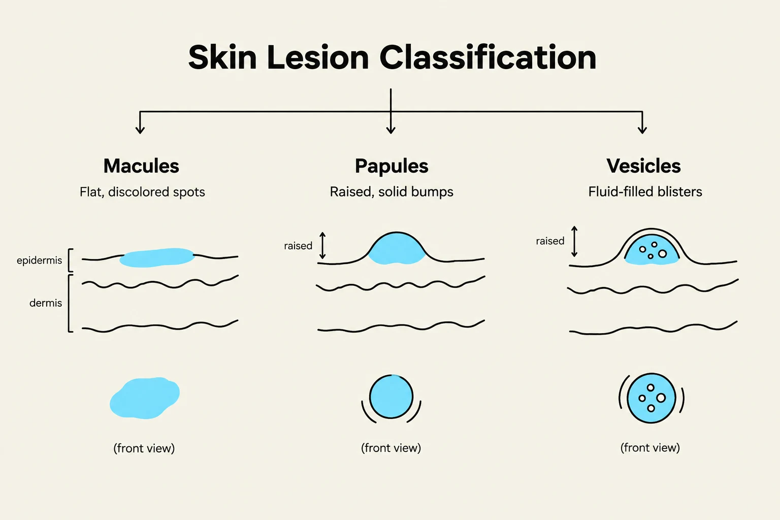
The first step in differential diagnosis is to move beyond the general term "rash" and describe the specific lesion type. A "rash" is a non-specific term that can encompass several distinct dermatological entities. To build an accurate differential, you must categorize the lesions based on their physical characteristics.
Key morphological descriptors include:
- Macules: Flat, non-palpable color changes in the skin.
- Papules: Small, raised, solid lesions.
- Vesicles: Small, fluid-filled blisters.
- Pustules: Small, pus-filled lesions.
- Erythema: Redness caused by increased blood flow to the superficial layers of the skin.
Exam Tip: Always check if the rash is "blanchable." Press on an erythematous lesion; if it turns white (blanches) and then returns to red, it is likely due to vasodilation. If it does not blanch, consider petechiae or purpura, which indicate bleeding into the skin.
Distribution and Pattern Recognition
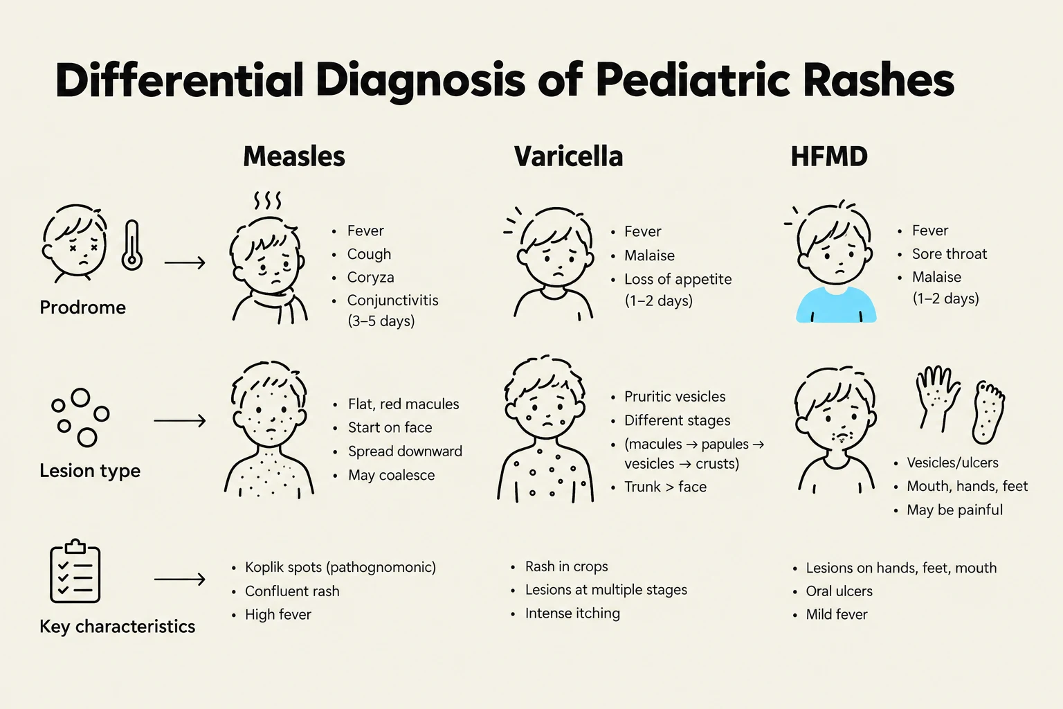
Once the morphology is established, the next critical question is: Where is the rash located? The distribution of a rash often provides the most significant clue to its etiology.
Keep reading for free.
Create an account to finish this reading and make it part of your study routine.
Continue reading freeAlready have an account? Log in
Updated