This content explains tendinopathy, a condition of persistent tendon pain related to mechanical loading. It covers the Cook-Purdam Model of pathophysiology, intrinsic and extrinsic risk factors, clinical diagnosis through history and physical examination, and treatment strategies including load management, progressive
Tendons are specialized collagenous tissues that connect muscle to bone, serving the critical function of transferring force from the muscular system to the skeletal system. While they possess excellent tensile properties, they are susceptible to injury through various mechanisms. When discussing overuse injuries, it is important to distinguish between enthesopathy—which specifically involves the injury or disorder of the enthesis (the junction where the tendon meets the bone)—and tendinopathy.
Tendinopathy is an umbrella term used to describe persistent tendon pain related to mechanical loading. Unlike acute injuries like ruptures, tendinopathy typically arises from repetitive tensile or compressive loading (such as sprinting or jumping) that causes repetitive microtraumas and inadequate recovery between loading periods.
The Pathophysiology: The Cook–Purdam Model
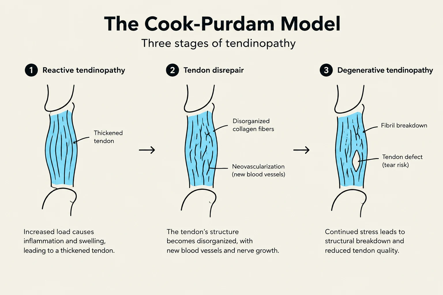
Understanding how a tendon progresses from healthy tissue to a chronic condition is essential for clinical management. The Cook–Purdam Model categorizes tendon pathology into three distinct stages:
Reactive tendinopathy: This is a non-inflammatory response where the tendon undergoes structural changes and thickening in the area being stressed.
Tendon disrepair: This represents a worsening of the pathology where the internal structure of the tendon becomes disorganized.
Degenerative tendinopathy: This is the chronic stage of the condition, often characterized by more significant structural breakdown.
The Cook-Purdam Model illustrates the three stages of tendinopathy: reactive tendinopathy, tendon disrepair, and degenerative tendinopathy, detailing the structural changes in tendons due to stress.
Clinical Note: It is recommended that clinicians use the term "tendinopathy" rather than "tendinitis" to avoid the implication that the condition is primarily an inflammatory process, as the reactive stage is often non-inflammatory.
Risk Factors
The development of tendinopathy is influenced by a combination of internal and external factors.
Intrinsic Risk Factors
These are characteristics inherent to the individual that may predispose them to injury:
Demographics: Older age, male sex, and menopause.
Biological/Systemic: Genetics, systemic conditions, and certain medications.
Physical: Biomechanics/technique and a history of previous injury.
Extrinsic Risk Factors
These are external variables related to the environment or training stimulus:
Loading Patterns: Total training load, sudden spikes in loads, and periods of deconditioning.
Changes: Sudden biomechanical changes in movement or equipment.
Clinical Diagnosis
Diagnosis is primarily clinical, relying on a thorough patient history and physical examination.
Patient History
A key diagnostic indicator is the progression of pain patterns. In many cases, the pain follows a predictable evolution:
Pain that appears only after exercise.
Pain that appears at the start of an activity.
Pain that occurs both during and after the activity.
Clinicians should also investigate the type of pain, as well as the patient's specific training and injury history.
Physical Examination and Imaging
The physical examination involves:
Inspection: Observing the area for swelling or visible changes.
Palpation: Identifying specific points of tenderness.
Range of Motion (ROM): Assessing the functional movement of the joint.
Pain Provocation Tests: Using specific movements to elicit the patient's pain.
While Ultrasound (US) and MRI are available and can be used to visualize the tendon, they are rarely required for a primary diagnosis.
This diagram illustrates the process of diagnosing tendinopathy, starting with patient history and progressing through physical examination (palpation and range of motion) to reach a diagnosis.
Treatment and Management Strategies
The primary goal of treatment is to manage the load on the tendon while promoting structural healing.
Load and Pain Management
Immediate management may include:
Load Management: Resting from the offending activity and utilizing orthoses.
Pain Control: Using ice, NSAIDs, and other passive treatments.
Progressive Strength Training
The cornerstone of long-term rehabilitation is progressive exercise therapy. For lower limb tendinopathy, rehabilitation typically follows a four-stage progression:
Stage
Exercise Type
Description
Stage 1
Isometric
Static holds to manage pain and maintain capacity
Stage 2
Isotonic & Heavy Slow Resistance (HSR)
Controlled movements through a range of motion
Stage 3
Energy Storage
Increasing speed and incorporating elastic loading
Stage 4
Energy Storage & Release
Sport-specific exercises and high-velocity movements
Recovery timelines vary significantly based on severity. Mild cases may resolve in 4–6 weeks, whereas moderate to severe cases may require 3–6 months. If surgery and subsequent rehabilitation are required, this timeline may extend to 6–7 months.
Other Modalities
In some clinical scenarios, other treatments may be considered, including shock wave therapy, laser, ultrasound, injectable therapies, or surgery.
Special Case: Rotator Cuff Tendinopathy
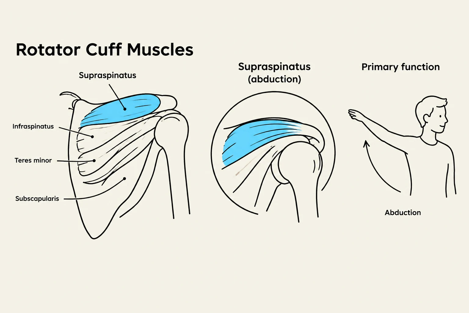
Tendinopathy of the shoulder often involves the four muscles that comprise the rotator cuff. Each muscle has a distinct functional role:
Supraspinatus: Responsible for abduction.
Subscapularis: Responsible for internal rotation.
Infraspinatus: Responsible for external rotation.
Teres Minor: Responsible for external rotation.
Management of shoulder injuries may also utilize clinical tools such as the OSTRC Shoulder Program for assessment and management.
Diagram illustrating the rotator cuff muscles and their role in shoulder abduction.
Exam Tip: When assessing shoulder pain, always differentiate which rotator cuff muscle is being tested based on the movement (e.g., testing external rotation helps identify potential infraspinatus or teres minor involvement).
Keep reading for free.
Create an account to finish this reading and make it part of your study routine.