An overview of Chronic Myeloid Leukemia (CML) and Acute Myelocytic Leukemia (AML), covering their distinct characteristics, diagnostic markers like the Philadelphia chromosome and Auer rods, and treatment approaches.
Leukemia and myeloproliferative disorders represent a group of malignant diseases characterized by the replacement of normal bone marrow with abnormal, neoplastic cells. This process results in a hypercellular bone marrow where the abnormal cells can invade the spleen, liver, lymph nodes, and peripheral blood. Understanding the distinction between chronic and acute forms, as well as the specific diagnostic markers used to differentiate similar conditions, is essential for clinical practice.
Chronic Myeloid Leukemia (CML)
Chronic Myeloid Leukemia (CML) is a clonal hematopoietic stem cell disorder characterized by the malignant transformation of a single stem cell. Clinically, patients often present with extreme leukocytosis (WBC counts often exceeding 100,000/μL), a severe "left shift" in the white cell differential, and splenomegaly.
The Philadelphia Chromosome
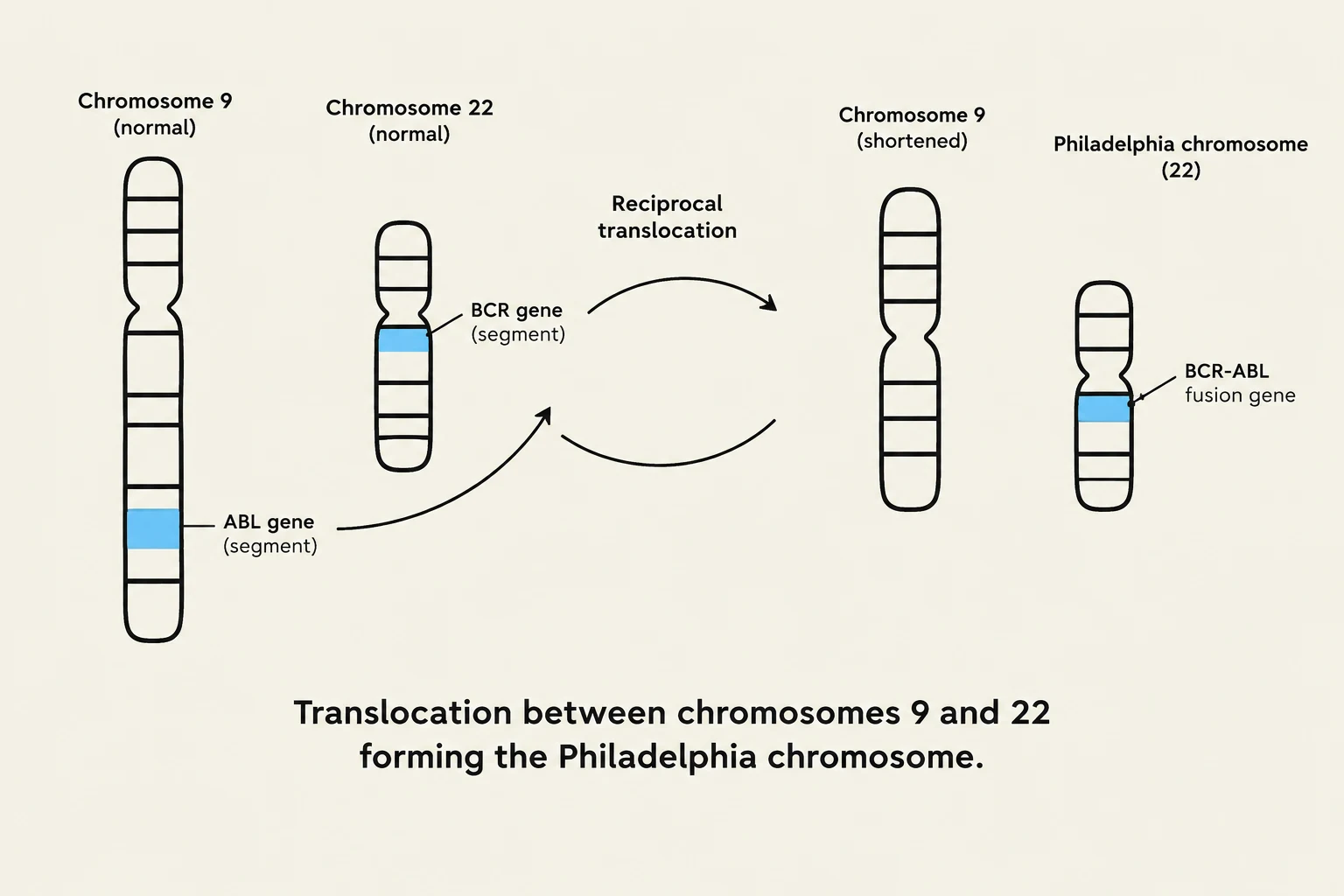
Diagram illustrating the reciprocal translocation between chromosomes 9 and 22, resulting in the formation of the Philadelphia chromosome and the BCR-ABL fusion gene.
A hallmark of CML is the presence of the Philadelphia Chromosome. This is a structural chromosomal abnormality resulting from a translocation between chromosomes 9 and 22, denoted as t(9;22). This translocation involves the BCR−ABL gene. Because this abnormality can only be demonstrated through cytogenetics, it serves as a primary diagnostic indicator. Interestingly, the presence of this chromosome can actually improve a patient's prognosis by making them more receptive to bone marrow transplants.
Distinguishing CML from Leukemoid Reaction
One of the most critical diagnostic challenges is differentiating CML from a leukemoid reaction—a high white blood cell count caused by infection or inflammation rather than malignancy. To make this distinction, clinicians utilize the Leukocyte Alkaline Phosphatase (LAP) score.
The LAP score measures the activity of alkaline phosphatase located in the specific granules of mature neutrophils. To calculate the score, 100 mature neutrophils are counted and graded on a scale of 0 to 4 based on their granular staining.
Diagnostic Category
LAP Score Range
Normal
40−170
Leukemoid Reaction
>190 (Increased)
CML
<20 (Decreased)
Exam Tip: Remember that CML is characterized by a low LAP score, whereas a leukemoid reaction (often due to infection) presents with an increased score. Note that these ranges can be subjective and may vary slightly between different laboratories.
Treatment and Progression
Treatment for CML typically involves chemotherapy, transplantation, and leukapheresis (a process used to remove excess white blood cells). Despite these interventions, many patients eventually progress into a blast crisis. During a blast crisis, the disease transforms into an acute leukemia that is often resistant to standard chemotherapy, significantly worsening the prognosis.
Acute Myelocytic Leukemia (AML)
While CML is a chronic disorder involving more mature cells, Acute Myelocytic Leukemia (AML) is characterized by a rapid progression and a high percentage of immature cells, known as blasts.
Diagnosis and Classification
Diagnosis of AML requires a bone marrow examination. Under the FAB (French-American-British) classification, a diagnosis is made if blasts constitute >30% of the bone marrow. The WHO classification is more modern, requiring >20% blasts if specific chromosomal abnormalities are present.
A hallmark finding in AML is the presence of Auer rods, which are bundles of primary granules that appear as pink or purple rods on a Wright stain.
The FAB system categorizes AML into several subtypes (M0–M7) based on morphology and cytochemical reactions:
M0: Minimally differentiated (no definitive myeloid features).
M1: Myeloblastic without maturation.
M2: Myeloblastic with maturation (often associated with t(8;21)).
M3: Promyelocytic (associated with t(15;17) and risk of DIC).
M4: Myelomonocytic.
M5: Monocytic.
M6: Erythroleukemia.
M7: Megakaryoblastic.
Cytochemical Staining
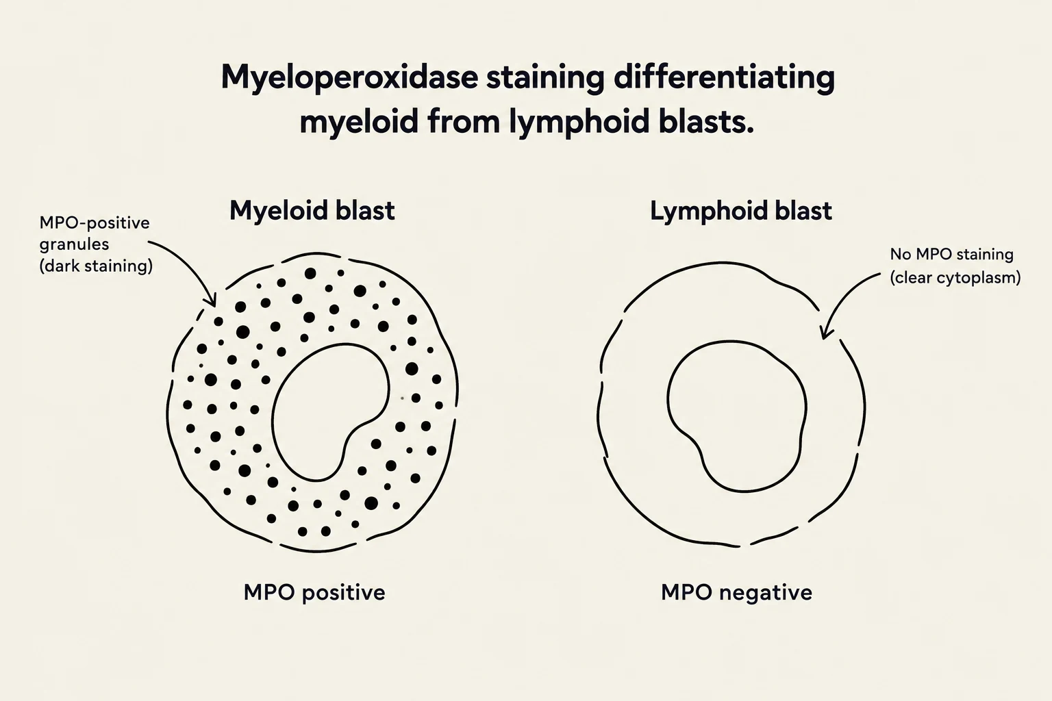
Diagram illustrating the difference between myeloid and lymphoid blasts based on myeloperoxidase (MPO) staining. Myeloid blasts show MPO-positive granules (dark staining), while lymphoid blasts lack MPO staining, resulting in clear cytoplasm.
To differentiate these subtypes, clinicians use special stains to identify specific enzymes or lipids in the leukemic cells:
Myeloperoxidase (MPO) & Sudan Black B: Used to differentiate myeloid blasts from lymphoid blasts.
Esterases: Used to identify monocytic origin. Non-specific esterase (NSE) is positive in monocytes (M4, M5), while Specific Esterase (SPE) is positive in myeloid cells.
Periodic Acid-Schiff (PAS): Helpful in diagnosing erythroleukemia (M6).
Treatment and Prognosis
The primary goal of AML treatment is to achieve remission, defined as the total absence of leukemic cells and the return of normal blood and bone marrow function.
Chemotherapy: Functions by blocking DNA or RNA synthesis. However, it is highly toxic and can lead to death via marrow hypoplasia.
Radiation: Often used in conjunction with chemotherapy.
Transplantation: This is considered the best chance for a true cure. The process involves "emptying" the marrow via chemo/radiation and then intravenously injecting autologous or HLA-matched marrow.
Clinical Note: Despite the potential of transplantation, it is high-risk; approximately $25-30%$ of patients die from rejection or infection.
The prognosis for AML in adults remains generally poor. While remission rates are between $45-75%$, remission typically lasts only 8−18 months. Only about $10%$ of adults survive past five years, though children and young adults generally experience significantly better outcomes.
Keep reading for free.
Create an account to finish this reading and make it part of your study routine.