


Respiratory Distress Syndrome (RDS), historically known as hyaline membrane disease, is a leading cause of morbidity in preterm infants. It is primarily driven by a deficiency in pulmonary surfactant, which leads to increased alveolar surface tension, widespread atelectasis, and impaired gas exchange. For clinicians and students, mastering the diagnostic approach—combining clinical observation, radiographic interpretation, and bedside physiological tests—is essential for timely intervention.
Radiographic Presentation: The 'Ground-Glass' Appearance
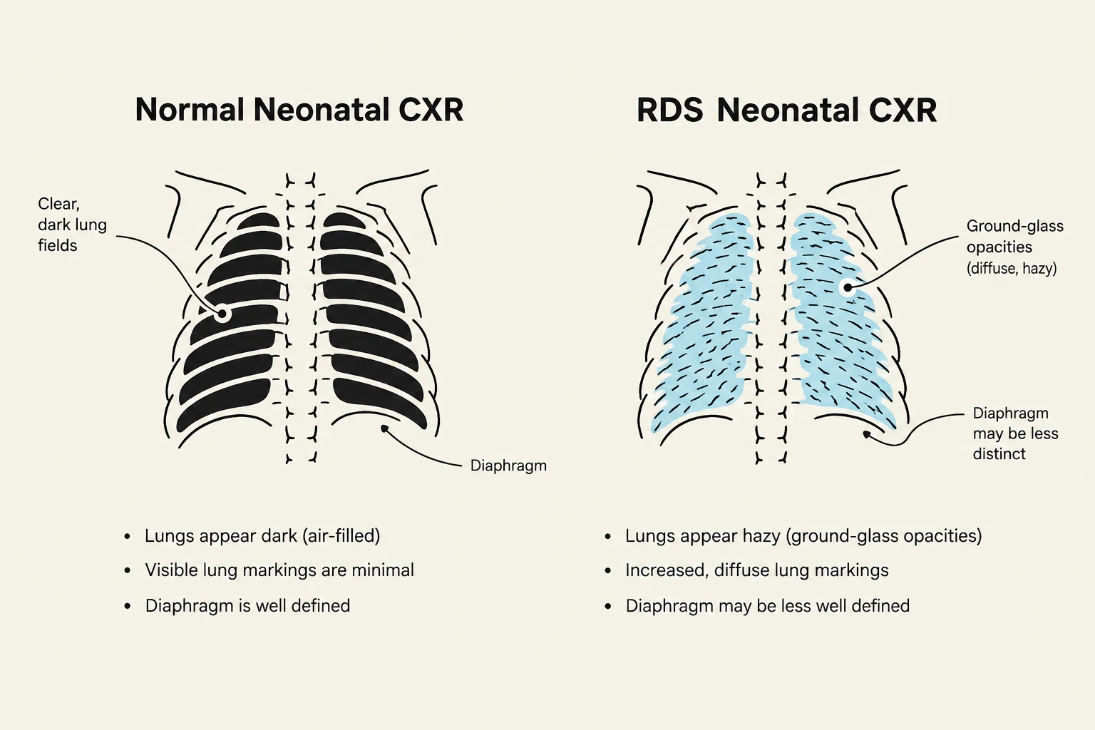
The gold standard for confirming a diagnosis of RDS in a clinical setting is the neonatal chest X-ray. Because the underlying pathology involves the collapse of small air sacs (alveoli) due to surfactant deficiency, the lungs do not appear "black" (air-filled) as they should in a healthy infant. Instead, they exhibit a characteristic pattern known as "ground-glass" opacification.
Ground-glass appearance refers to a fine, granular, or hazy pattern of increased density that covers the lung fields. This occurs because the alveoli are partially collapsed or filled with proteinaceous material, preventing the X-ray beams from passing through freely. On a radiograph, this looks like a diffuse, symmetric "mist" or "sand-like" texture across both lungs.
In addition to this hazy texture, clinicians should look for:
- Air bronchograms: As the alveoli collapse and become opaque, the air-filled bronchi (the tubes leading to the alveoli) become visible as dark, branching lines against the white, "ground-glass" background.
- Low lung volumes: Because the lungs are difficult to inflate without surfactant, the overall volume of air in the chest appears reduced compared to a healthy neonate.
Keep reading for free.
Create an account to finish this reading and make it part of your study routine.
Continue reading freeAlready have an account? Log in
Updated