


Ossification, also known as osteogenesis, is the biological process of bone formation. This process is not a single event but occurs in several distinct scenarios throughout a human life cycle: during pre-natal development in the embryo and fetus, during the adolescent growth period from infancy to adulthood, during the continuous remodeling of adult bone, and during the repair of fractures.
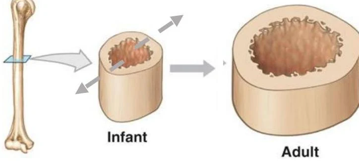
Understanding how bones grow requires distinguishing between two primary mechanisms: how they get thicker and how they get longer.
Pre-Natal Bone Formation: Two Pathways
Before birth, the skeletal system begins to form through two different methods depending on the type of bone being created.
Intramembranous Ossification
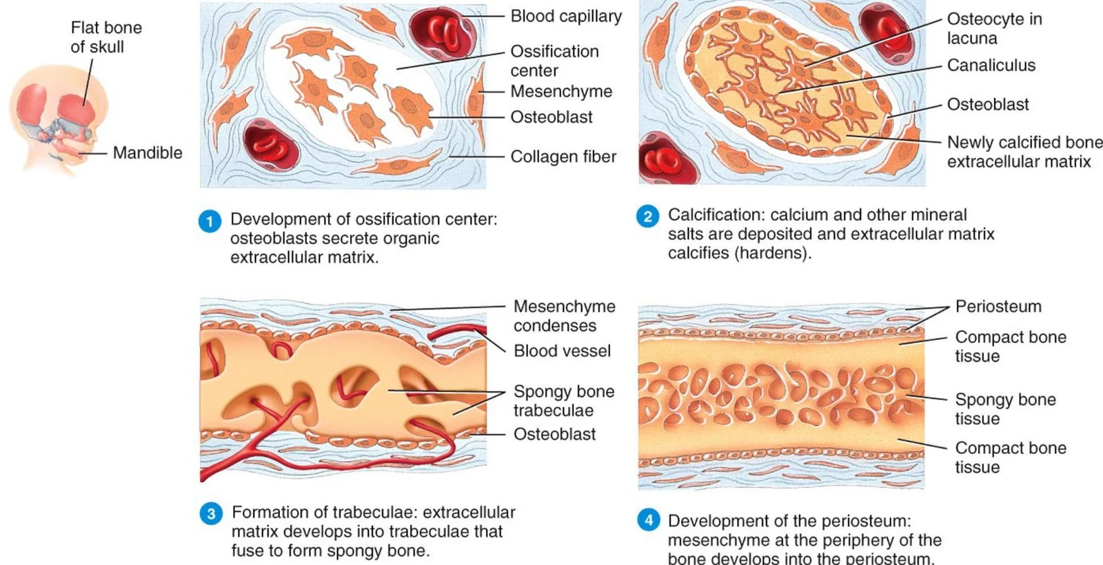
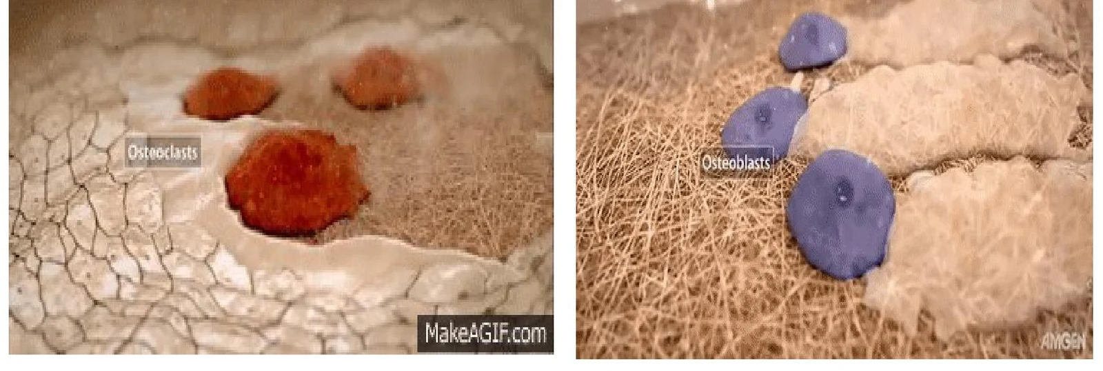
This is a simpler method used primarily for the formation of flat bones, such as those in the skull and the clavicle. The process begins when mesenchymal cells develop into osteoprogenitor cells, which then differentiate into osteoblasts. These osteoblasts form an ossification center by secreting an organic extracellular matrix. As calcium and other mineral salts are deposited, this matrix calcifies and hardens. The developing bone tissue eventually organizes into trabeculae, forming spongy bone, while the mesenchyme at the periphery develops into the periosteum.
Endochondral Ossification
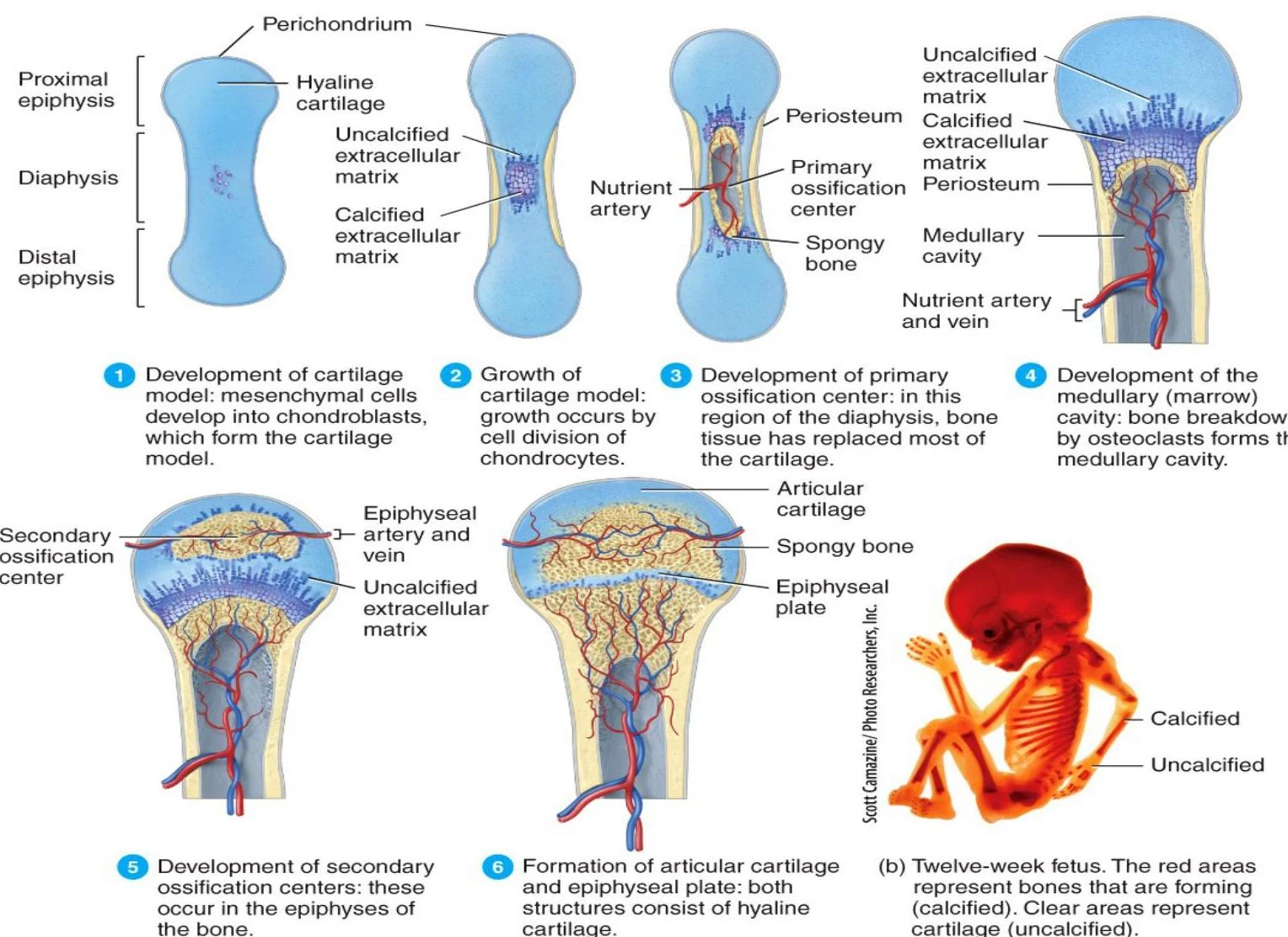
Most bones, particularly long bones, form through endochondral ossification, which involves the replacement of a cartilage model with bone tissue.
The process follows a specific sequence:
- Cartilage Model Development: Mesenchymal cells become chondroblasts, which create a cartilage model.
- Growth of the Model: The model grows through the cell division of chondrocytes.
- Primary Ossification Center: In the region of the diaphysis (the shaft of the bone), bone tissue replaces most of the cartilage.
- Medullary Cavity Formation: Osteoclasts break down bone to create the marrow cavity.
- Secondary Ossification Centers: These develop in the epiphyses (the ends of the bone).
Keep reading for free.
Create an account to finish this reading and make it part of your study routine.
Continue reading freeAlready have an account? Log in
Updated