


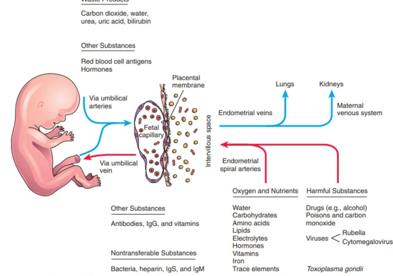
In the early stages of embryogenesis, the developing embryo relies on simple diffusion to receive nutrients and oxygen. However, as the fetus grows, its metabolic demands increase significantly, surpassing the effective distance for simple diffusion (approximately ). To sustain life, a complex organ called the placenta develops, creating a massive surface area to facilitate efficient gaseous and nutrient exchange between the maternal and fetal components.
The placenta is a dual-origin organ, meaning it is composed of both maternal and fetal tissues. The maternal portion is derived from the decidua basalis, while the fetal portion originates from the trophoblast.
Trophoblast Differentiation and Implantation
During the process of implantation, the trophoblast differentiates into two distinct layers that are fundamental to placental architecture:
- Cytotrophoblast: The inner layer consisting of individual cells.
- Syncytiotrophoblast: The outer layer, which forms a continuous, multinucleated mass known as a syncytium.
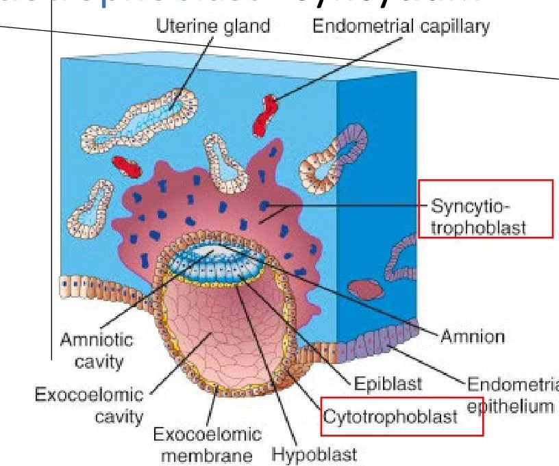
As the embryo develops, the extraembryonic (EE) coelom forms the chorionic cavity, and the structures forming the outer border of this cavity are collectively referred to as the chorion. The chorion is composed of the somatopleuric extraembryonic mesoderm, the cytotrophoblast, and the syncytiotrophoblast.
Development of the Chorionic Villi
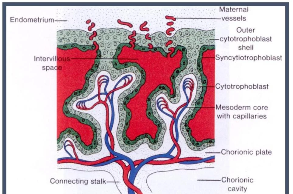
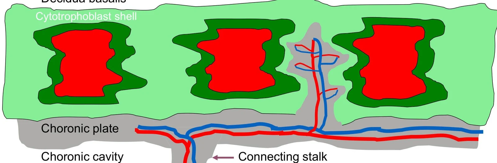
The functional unit of the fetal placenta is the chorionic villus. These villi develop from trabeculae (spaced columns) that form within the lacunae. The development of these villi occurs in stages:
- Primary Villus: Consists of a core of cytotrophoblast cells covered by a layer of syncytiotrophoblast.
- Secondary Villus: (Implied by the progression of mesoderm penetration).
- Tertiary Villus: These are the mature villi. They are characterized by the penetration of extraembryonic (EE) mesoderm into the villus core, which eventually gives rise to fetal blood vessels.
Keep reading for free.
Create an account to finish this reading and make it part of your study routine.
Continue reading freeAlready have an account? Log in
Updated