A comprehensive review of dental anatomy, including oral tissues, tooth structure, morphology, development, eruption, and occlusion classifications. Covers concepts like tooth numbering systems, primate spacing, and malocclusion types.
Preparing for your first anatomy exam requires a systematic approach to understanding how the mouth is structured, how teeth are built, and how they interact during function. This review guide organizes the core concepts of oral anatomy, tooth morphology, development, and occlusion to help you navigate the complexities of the ANAT 1119 curriculum.
Oral Anatomy and Mucosal Tissues
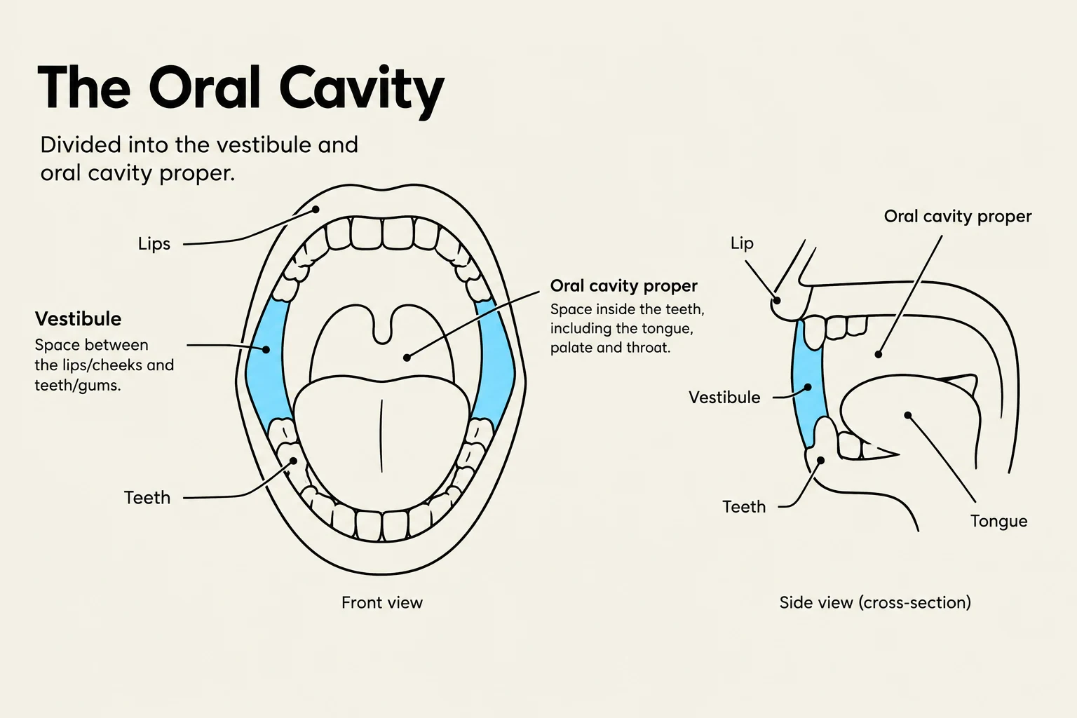
The oral cavity is not a uniform space; it is divided into two primary regions: the vestibule (the space between the lips/cheeks and the teeth) and the oral cavity proper (the area inside the teeth).
The mucosal tissues lining these areas vary significantly based on their function and location. You must be able to distinguish between:
Labial mucosa: Lines the inner surface of the lips.
Buccal mucosa: Lines the inner surface of the cheeks.
Alveolar mucosa: The loose, red tissue covering the alveolar bone.
Attached gingiva: The firm, pink tissue that is tightly bound to the underlying bone.
The boundary where the movable alveolar mucosa meets the firm attached gingiva is known as the mucogingival junction. While studying these tissues, you may encounter "variations of normal," such as mandibular tori—bony outgrowths on the lingual surface of the mandible that are considered normal anatomical variations rather than pathologies.
Diagrams illustrating the front and side views of the oral cavity, labeling its main parts such as lips, teeth, vestibule, and oral cavity proper.
Tooth Structure and Morphology
A tooth is divided into two main parts: the crown and the root. It is vital to distinguish between the anatomical crown (the part covered by enamel) and the clinical crown (the part of the tooth visible in the mouth). Vertically, the tooth is further divided into the coronal (crown), cervical (neck), and apical (root tip) portions.
Tooth Tissues
The tooth is composed of four primary hard and soft tissues:
Enamel: The highly mineralized, hardest tissue covering the crown.
Dentin: The bulk of the tooth structure located beneath the enamel and cementum.
Cementum: The bone-like tissue covering the root.
Pulp: The soft tissue in the center containing nerves and blood vessels.
The Cementoenamel Junction (CEJ) is a critical landmark, as it marks the anatomical boundary where the enamel of the crown meets the cementum of the root.
Tooth Types and Surfaces
Teeth are categorized by their function:
Incisors: Used for cutting.
Canines: Used for tearing.
Premolars: Used for crushing/piercing.
Molars: Used for grinding.
To describe a tooth's location, we use specific surface terminology: labial or buccal (the outer surface), lingual (the inner surface), mesial (the side toward the midline), distal (the side away from the midline), occlusal (the biting surface of posterior teeth), and incisal (the biting edge of anterior teeth).
Exam Tip: When identifying surfaces, always orient yourself to the midline of the dental arch. "Mesial" always moves toward the center, and "Distal" always moves toward the back of the mouth.
Tooth Identification and Development
Numbering Systems
In clinical practice, you must be able to convert between numbering systems. A common exam question involves the FDI system (which uses two digits) and the Universal system.
Example Conversion:
If a tooth is identified as 11 in the FDI system, it corresponds to tooth 8 in the Universal system.
Development and Eruption
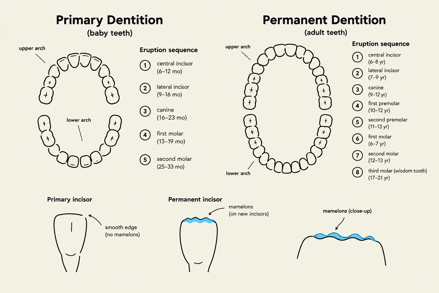
Humans transition from primary dentition (baby teeth) to permanent dentition (adult teeth). During the formation of anterior teeth, you may observe mamelons—small, rounded protuberances on the incisal edges of newly erupted permanent incisors. In the posterior teeth, specifically molars, you may see the Cusp of Carabelli, a supplemental cusp found on maxillary first molars.
Understanding eruption dates is essential. If a primary tooth is lost prematurely, it can lead to space loss and malocclusion, as the surrounding teeth may drift into the empty space.
This illustration compares primary (baby) and permanent (adult) dentition, detailing the eruption sequence and characteristics of each type of tooth.
Occlusion and Space Management
Occlusion refers to how the upper and lower teeth meet. This is categorized into dental occlusion (the relationship of the teeth) and skeletal occlusion (the relationship of the jaw bones).
Space Concepts
Primate spacing: Natural gaps found in the dental arches of children.
Leeway spacing: The difference in size between the primary molars/canines and the permanent premolars/canines that replaces them. This extra space is crucial for the proper alignment of permanent teeth.
Occlusal Relationships and Classifications
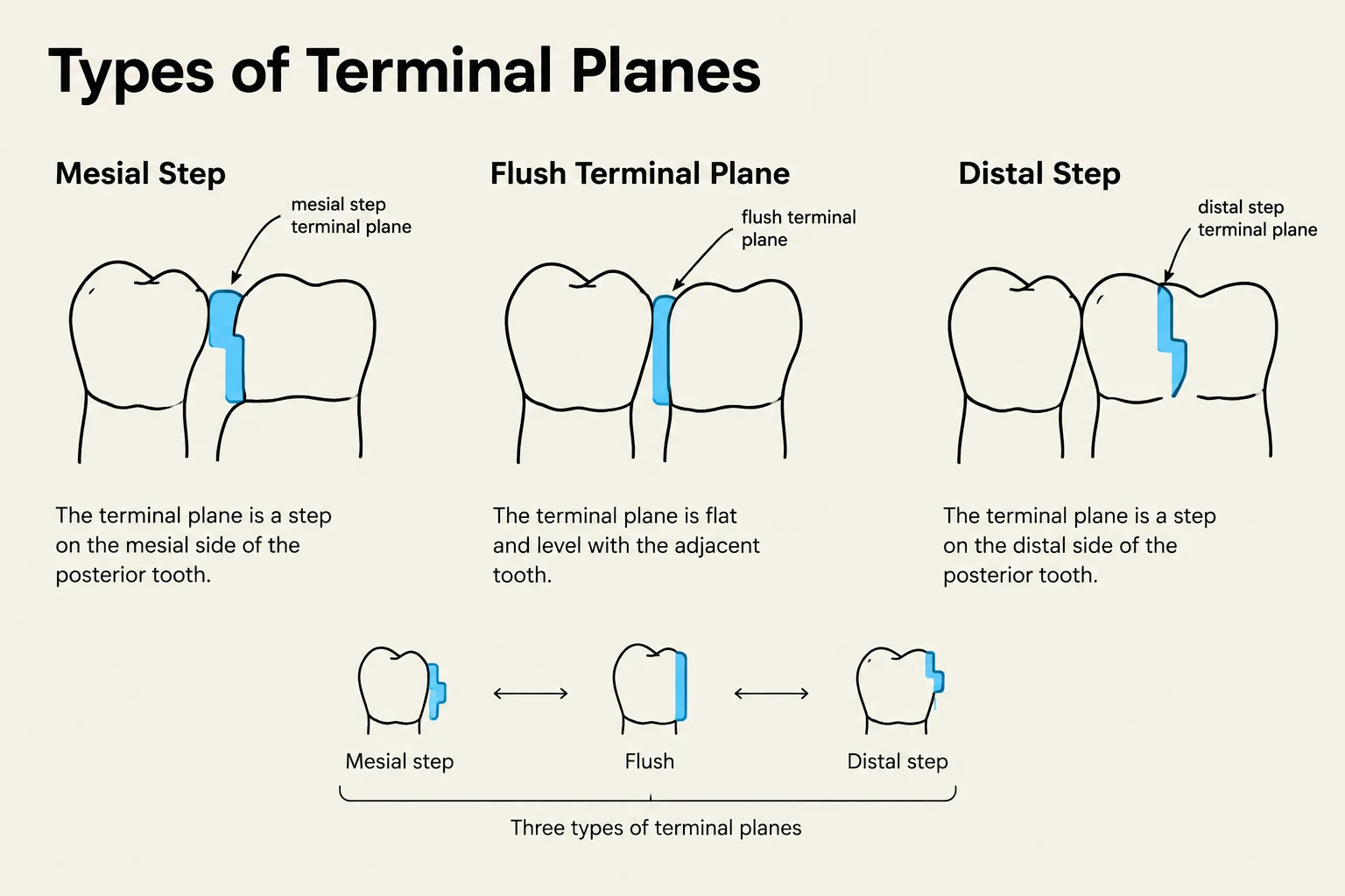
To classify occlusion, clinicians look at the position of the permanent first molars and the terminal planes (the relationship of the most distal primary molars):
Mesial step: The mandibular molar is mesial (forward) to the maxillary molar.
Flush terminal plane: The surfaces of the distalities are even.
Distal step: The mandibular molar is distal (behind) to the maxillary molar.
The major classifications are:
Class I: Normal relationship.
Class II: The maxillary teeth are positioned too far forward relative to the mandibular teeth (often associated with overjet, the horizontal overlap).
Class III: The mandibular teeth are positioned too far forward (often associated with an increased overbite, the vertical overlap).
Note on Malocclusion:
Deep bite: Excessive vertical overlap.
Open bite: A lack of vertical overlap.
Cross bite: When upper teeth sit inside the lower teeth.
Functional Concepts
Finally, distinguish between centric occlusion (the position of the teeth when they are fully biting together) and centric relation (the relationship of the mandible to the maxilla when the condyles are in their most superior-anterior position in the glenoid fossa).
Diagram illustrating the three types of terminal planes in dentistry: Mesial Step, Flush Terminal Plane, and Distal Step.
Keep reading for free.
Create an account to finish this reading and make it part of your study routine.