Dehydration: Causes, Assessment, and Electrolyte Dynamics
This content explains dehydration as a fluid volume deficit, detailing its causes, the body's compensatory cardiovascular and neurological responses, and the resulting electrolyte and hematological changes.
Dehydration, clinically referred to as a fluid volume deficit, is a state where the body's fluid loss exceeds its intake. This disruption is rarely a sudden, isolated event; rather, it is often a cumulative process that progressively undermines the body's metabolic and homeostatic balance. Because water and electrolytes are inextricably linked—water often moves in response to the movement of solutes—dehydration can trigger a cascade of physiological shifts affecting everything from blood pressure to neurological function.
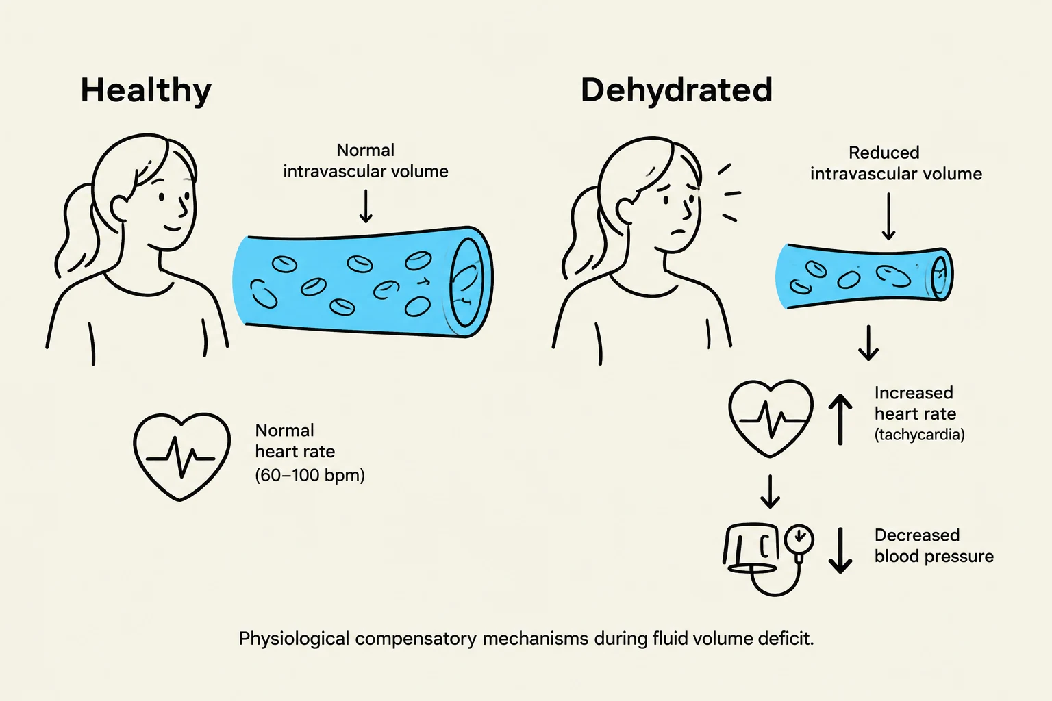
This illustration depicts the physiological compensatory mechanisms during fluid volume deficit, comparing a healthy state with a dehydrated state. In dehydration, reduced intravascular volume leads to increased heart rate (tachycardia) and decreased blood pressure.
Causes of Dehydration
The drivers of dehydration can be categorized into increased loss, reduced intake, and acute volume depletion.
Increased Fluid Loss
Common physiological drivers include vomiting and diarrhea, which directly remove both water and electrolytes from the body. Fever is another significant factor; as the body's metabolic demands ramp up to generate heat, fluid loss increases. Additionally, medical interventions and chronic conditions such as the use of diuretics or uncontrolled diabetes can lead to persistent fluid depletion.
Reduced Intake
A lack of sufficient intake is a major contributor, particularly in vulnerable populations. A critical factor in the elderly is hypodipsia, a diminished sensation of thirst that prevents them from recognizing the need for fluids. Cognitive impairments—such as sedation, delirium, or depression—can also prevent a patient from seeking hydration. Furthermore, some patients may intentionally limit their intake due to mobility issues or a fear of urinary incontinence.
Acute Loss and Hemorrhage
Significant blood loss, such as a major hemorrhage or even large hematomas (bruising), constitutes a form of dehydration. This is because blood loss involves the loss of plasma, which is the liquid component of the intravascular space.
Assessment and Monitoring
Recognizing dehydration requires a multi-system approach, as the body attempts to compensate for the shrinking intravascular volume.
Cardiovascular Responses
The cardiovascular system is often the first to show signs of a fluid volume deficit. As the circulating blood volume drops, the heart attempts to maintain cardiac output by increasing the heart rate. This can lead to:
Dysrhythmias: Irregular heart rhythms due to electrolyte shifts and stress.
Weak Peripheral Pulses: Reduced pressure in the extremities.
Orthostatic Hypotension: A significant drop in blood pressure when moving from a lying to a sitting or standing position.
Syncope: In severe cases, the inability to maintain cerebral perfusion can lead to fainting.
Vascular and Neurological Signs
Clinicians must look beyond blood pressure. While Jugular Venous Distention (JVD) is a hallmark of fluid excess, the presence of flat veins can indicate dehydration. As dehydration progresses, neurological decline may occur, manifesting as altered mental status or diminished deep tendon reflexes.
Effective Monitoring Tools
To track fluid status accurately, healthcare providers use several methods:
"Ins and Outs" (I&O): Measuring total fluid intake against urine output, wound drainage, and perspiration.
Daily Weights: For chronic patients, this is the most effective monitoring method. To ensure accuracy, weights should be taken at the same time, on the same scale, and with similar clothing.
Clinical Tip: When assessing mucous membranes, check the tongue and the underside of the eyelid. If they are dry rather than moist and pink, it is a strong indicator of dehydration.
Electrolyte and Hematological Changes
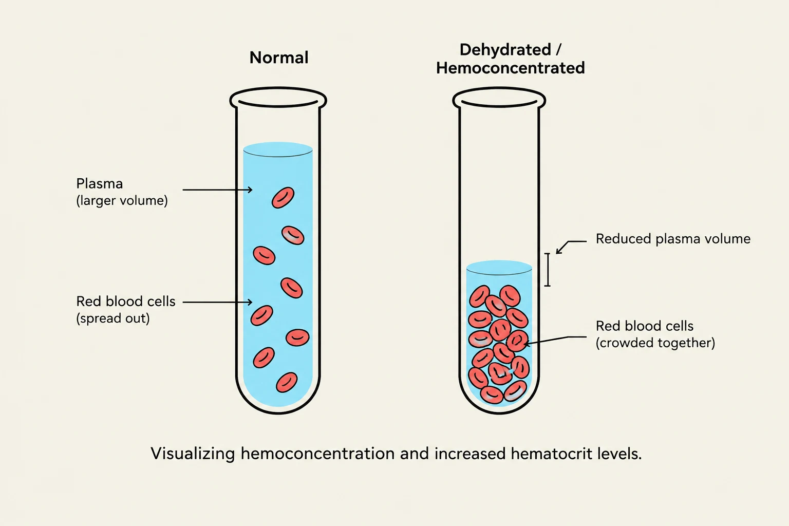
This diagram illustrates the difference between normal and dehydrated/hemoconcentrated blood samples, showing reduced plasma volume and crowded red blood cells in the latter.
Dehydration does not always result in low electrolyte levels; the concentration depends on whether the patient has lost "free water" or a combination of water and solutes.
Electrolyte Concentration Dynamics
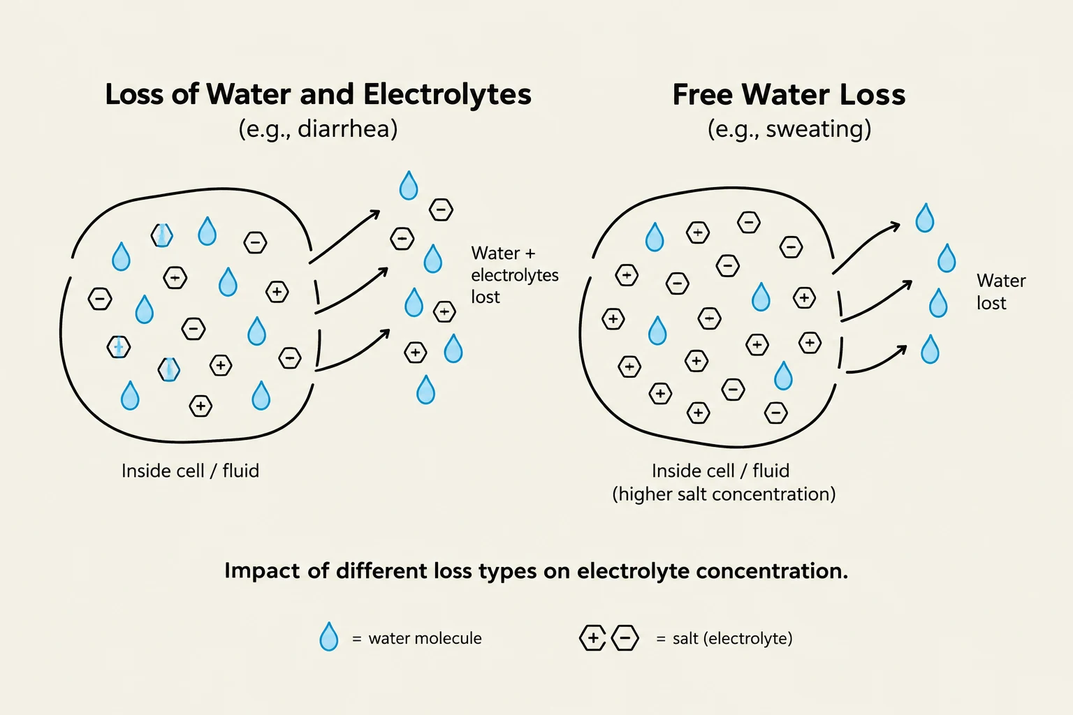
Decreased Electrolytes: This occurs when electrolytes are lost alongside water, such as during heavy sweating, vomiting, or diarrhea.
Increased Electrolytes: This occurs during free water loss. If a patient loses water but retains solutes like sodium, potassium, and chloride, the remaining plasma becomes more concentrated. This leads to elevated lab values for these electrolytes.
Exam Alert: Laboratory values obtained via venipuncture reflect the concentration in the plasma only. They may not accurately represent the fluid status in other compartments, such as the intracellular space.
Hemoconcentration
As plasma volume decreases, the blood becomes more concentrated. This is measured via the hematocrit, which is the percentage of red blood cells (RBCs) in the total blood volume.
Consider this example of hemoconcentration:
In a normal state, a patient might have a hematocrit of 45%. If the patient loses 15% of their plasma volume through dehydration, the number of red blood cells remains the same, but they are now squeezed into a smaller amount of fluid. The mathematical relationship can be viewed as:
Hematocrit=Total Blood Volume (Plasma + RBCs)Volume of RBCs
As the denominator (Total Blood Volume) decreases due to plasma loss, the resulting hematocrit percentage increases, even though no new red blood cells were created.
Osmolality and Specific Gravity
This diagram illustrates the difference between loss of water and electrolytes (e.g., diarrhea) and free water loss (e.g., sweating) on electrolyte concentration within a cell or fluid.
Because dehydration involves higher concentrations of solutes, both serum osmolality and urine specific gravity will typically be elevated. The body attempts to conserve water, resulting in highly concentrated urine.
Summary Table: Fluid Volume Deficit Indicators
System
Early/Mild Signs
Late/Severe Signs
Cardiovascular
Increased heart rate, orthostatic hypotension
Dysrhythmias, syncope, weak pulses
Neurological
Mild confusion, thirst
Altered mental status, diminished reflexes
Integumentary
Dry mucous membranes
Skin tenting (decreased turgor)
Laboratory
Increased hematocrit, increased urine specific gravity
Elevated sodium/potassium (if free water loss)
Keep reading for free.
Create an account to finish this reading and make it part of your study routine.