Definitions and Mechanisms of Pain and Nociception
An overview of the physiological processes of nociception, sensory fiber types, and the neurobiological pathways involved in pain perception, including transduction, conduction, transmission, modulation, and perception.
Understanding the difference between the sensation of pain and the physiological process of nociception is fundamental to clinical pharmacology and neurology. While often used interchangeably in casual conversation, they represent distinct biological phenomena: one is a physiological mechanism, and the other is a subjective experience.
Defining Pain and Nociception
The International Association for the Study of Pain defines pain as an "unpleasant sensory and emotional experience associated with actual or potential tissue damage or described in terms of such damage". This definition highlights that pain is not merely a physical signal; it includes an affective (emotional) component that can involve anxiety, fear, or suffering.
In contrast, nociception is the physiological neural process of perceiving noxious stimuli. It is the mechanical, thermal, or chemical detection of a stimulus by specialized neurons, which then converts that stimulus into an electrical signal.
Exam Tip: Remember that nociception is the process (the "hardware" and "software" running the signal), while pain is the perception (the "user experience"). You can have nociception without the conscious perception of pain, but pain is always driven by nociceptive processes.
The Five Stages of Nociception
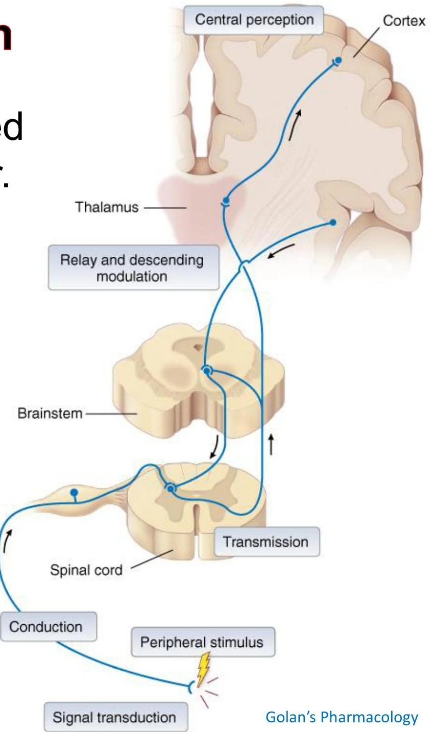
The journey of a pain signal from a stimulus to the brain follows a specific sequence of five stages: transduction, conduction, transmission, perception, and modulation.
Diagram illustrating the pathway of sensory information from a peripheral stimulus to the brain, involving the spinal cord, brainstem, thalamus, and cortex, with relay and descending modulation.
1. Transduction and Peripheral Sensitization
Transduction occurs at the site of injury via nociceptors, which are unspecialized free nerve endings. These receptors convert noxious stimuli into action potentials. The intensity of the stimulus is encoded by the frequency of the action potentials generated.
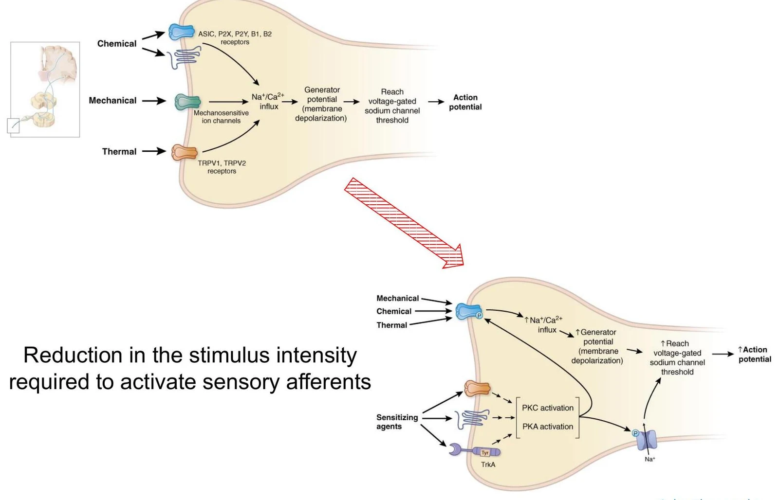
Different receptors handle different types of stimuli:
ASIC (Acid-Sensing Ion Channels): Respond to low pH (high $H^+$ concentration).
P2X: Ligand-gated ion channels that respond to ATP.
B1/B2 Receptors: G protein-coupled receptors that respond to kinin peptides.
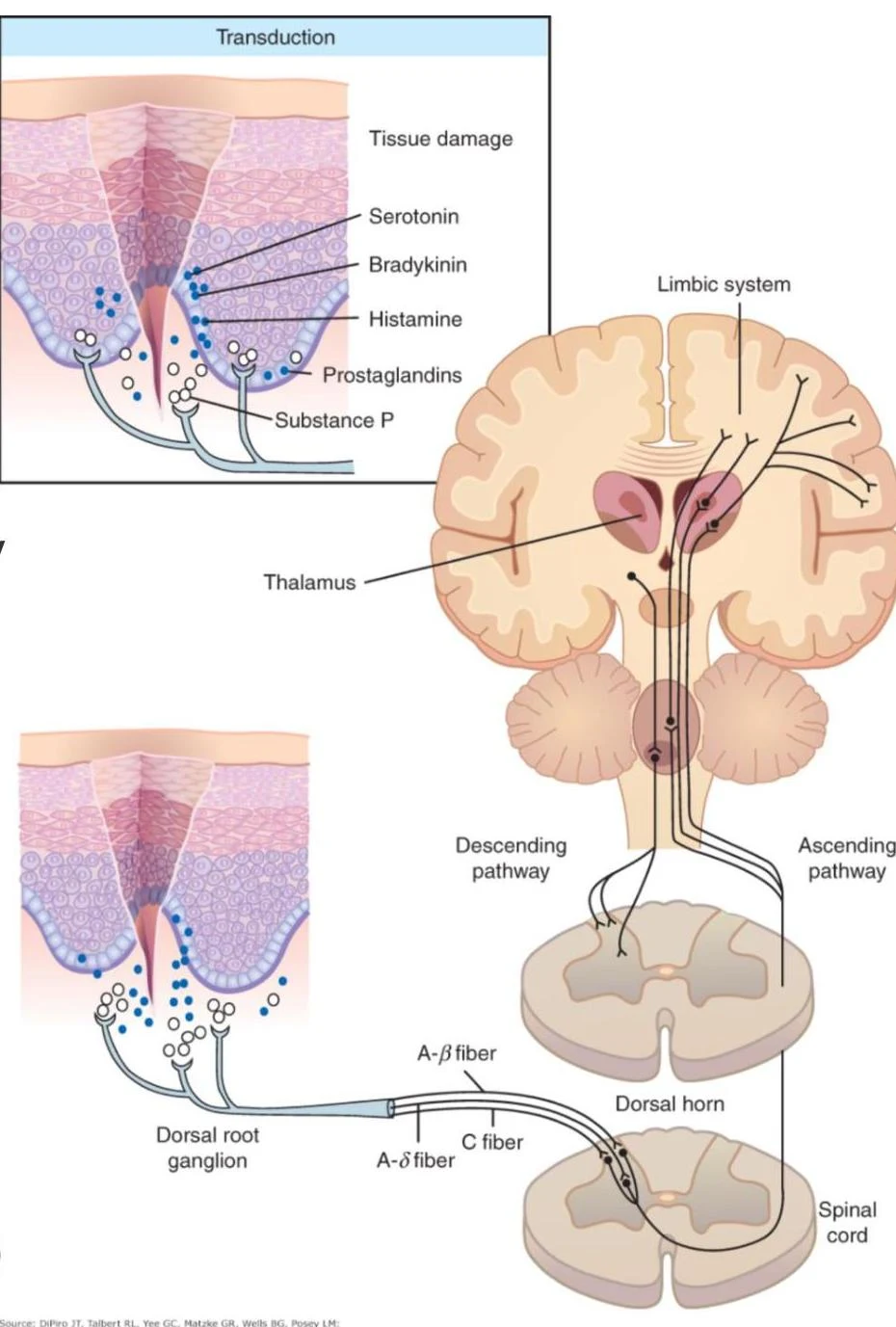
When tissue is injured, it releases a "soup" of chemical mediators, including bradykinin, substance P, and various prostaglandins. Prostaglandins (specifically PGE2 and PGI2) play a critical role by increasing the sensitivity of the nerve terminals. This process is known as peripheral sensitization, where the threshold required to activate the sensory afferents is lowered, making the area more sensitive to even mild stimuli.
Diagram illustrating the process of sensory afferent activation and how sensitizing agents can reduce the stimulus intensity required for activation, leading to increased neuronal excitability.
2. Conduction
Once the signal is transduced, it travels along peripheral nerve fibers via conduction. There are two primary types of fibers involved in transmitting pain signals, each with distinct characteristics:
Feature
Aδ (A-delta) Fibers
C Fibers
Myelination
Thinly myelinated
Unmyelinated
Conduction Speed
Fast (5−25 m/s)
Slow (0.5−2.3 m/s)
Pain Type
"First pain": Sharp, localized
"Second pain": Dull, diffused
Neurochemistry
Primarily Glutamate
Glutamate, Substance P, CGRP
Diagram illustrating a peripheral nerve connecting to the spinal cord, including the dorsal root ganglion and sympathetic preganglionic neuron.
3. Transmission
Transmission involves the movement of the signal from the peripheral nervous system into the central nervous system. In acute, protective pain, the signal travels via the Spinothalamic Tract (STT).
The pathway follows these steps:
The primary afferent neuron enters the spinal cord and synapses in the dorsal horn.
The second-order neuron decussates (crosses over) to the contralateral side of the spinal cord.
The signal ascends through the spinothalamic tract to the thalamus.
The third-order neuron carries the signal from the thalamus to the somatosensory cortex.
This diagram illustrates the process of pain transduction, showing how tissue damage leads to the release of neurotransmitters like serotonin, bradykinin, histamine, prostaglandins, and substance P. These signals are transmitted via nerve fibers (A-beta, A-delta, and C fibers) through the dorsal root ganglion and spinal cord to the thalamus and limbic system in the brain.
Central Sensitization: Hyperalgesia and Allodynia
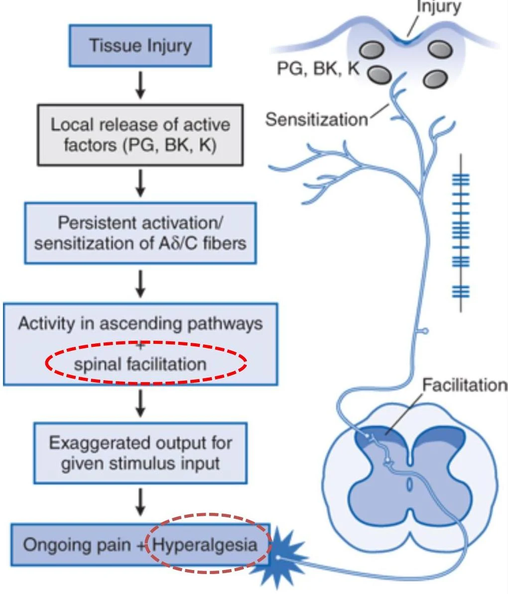
If nerve injury occurs (e.g., from diabetes or shingles), the continuous influx of signals can lead to central sensitization. This is a state of spinal facilitation where the spinal neurons become hyper-responsive. This results in two clinical phenomena:
Hyperalgesia: An exaggerated, increased pain response to a stimulus that is normally painful.
Allodynia: Pain caused by a stimulus that is normally not painful (innocuous), such as a light touch.
This diagram illustrates the physiological process of hyperalgesia, which begins with tissue injury and leads to increased sensitivity to pain.
4. Modulation
The body does not passively receive pain signals; it actively attempts to dampen them through descending inhibitory pathways. These pathways originate in the brain and travel down to the dorsal horn of the spinal cord to suppress nociceptive input.
Key neurochemical players in this descending control include:
Locus Coeruleus: Uses epinephrine (norepinephrine).
These descending signals can act directly on nociceptive neurons or via inhibitory interneurons to release GABA or opioids, effectively "closing the gate" on the pain signal before it reaches the brain.
Diagram illustrating the noradrenergic and serotonergic pathways originating in the brainstem and descending to the spinal cord.
5. Perception
The final stage is perception, where the signal is integrated in the brain. This involves the thalamocortical projections to the somatosensory cortex (which localizes the pain) and the limbic system (which processes the emotional and affective dimensions).
Clinical Phenomenon: Referred Pain
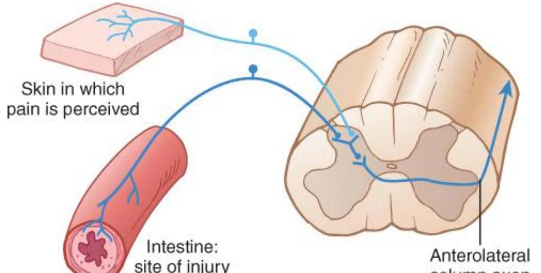
Sometimes, pain is felt in a location far from the actual site of injury. This is known as referred pain. This occurs because cutaneous (skin) sensory neurons and visceral (organ) sensory neurons often converge onto the same second-order projection neuron in the spinal cord's dorsal horn. When the brain receives a signal from that second-order neuron, it may misinterpret the source as coming from the skin rather than the internal organ.
This diagram illustrates the pathway of pain signals from the intestine to the spinal cord and then up the anterolateral column.
Diagrams illustrating referred pain patterns in the human torso, showing areas of pain associated with internal organs.
Keep reading for free.
Create an account to finish this reading and make it part of your study routine.