Definitions and Mechanisms of Pain and Nociception
This document explains the distinction between pain and nociception, detailing the five stages of nociception: transduction, conduction, transmission, perception, and modulation. It also covers nerve injury, abnormal pain responses like hyperalgesia and allodynia, and the phenomenon of referred pain.
To the casual observer, pain is a simple reaction to injury. However, in the realms of neurobiology and pharmacology, pain is recognized as a complex, multi-dimensional experience that involves distinct physiological processes and profound emotional components. Understanding the difference between the physical detection of a stimulus and the subjective experience of suffering is fundamental to treating everything from a minor burn to chronic neuropathic conditions.
Defining Pain vs. Nociception
It is crucial to distinguish between pain and nociception. The International Association for the Study of Pain defines pain as an "unpleasant sensory and emotional experience associated with actual or potential tissue damage or described in terms of such damage". This definition highlights that pain is not merely a physical signal; it includes an emotional and psychological dimension that can exist even in the absence of visible tissue damage.
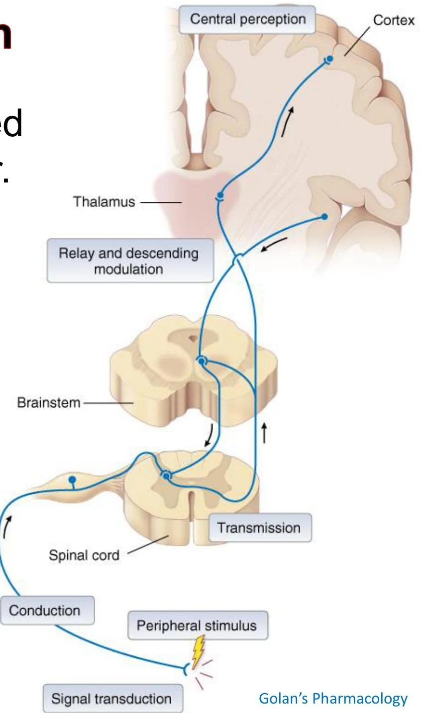
Nociception, by contrast, is the purely physiological process. It refers to the neural mechanism by which the body perceives noxious (harmful) stimuli through a specific sequence of stages: transduction, conduction, transmission, perception, and modulation.
Diagram illustrating the pathway of sensory information from a peripheral stimulus to the brain, involving the spinal cord, brainstem, thalamus, and cortex, with relay and descending modulation.
The Five Stages of Nociception
The journey from a physical stimulus to a conscious sensation follows a highly organized pathway.
1. Transduction and Peripheral Sensitization
Transduction is the first step, occurring at the site of injury. Nociceptors—unspecialized sensory neurons with free nerve endings—act as transducers. They convert mechanical, thermal, or chemical energy into electrical signals known as action potential (AP) trains. The frequency of these action potentials is directly proportional to the intensity of the stimulus.
Various receptors are specialized for different stimuli:
ASIC (Acid-Sensing Ion Channels): Respond to low pH (high H+ concentration).
P2X receptors: Ligand-gated ion channels that respond to ATP.
B1 and B2 receptors: G protein-coupled receptors that respond to kinin peptides.
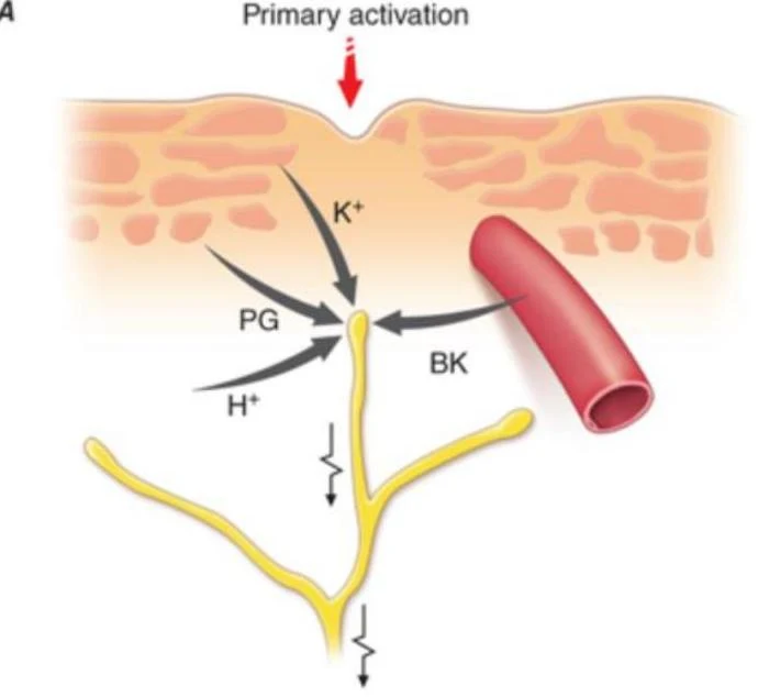
When tissue is injured, a "chemical soup" is released, including prostaglandins (PGE2 and PGI2), bradykinin, and substance P. These mediators cause peripheral sensitization, a process where the threshold for activating nociceptors is lowered. This means a stimulus that was previously painless may now trigger a response, and the intensity of the pain is amplified.
Diagram illustrating the primary activation of a nerve ending by various chemical mediators including potassium ions (K+), prostaglandins (PG), bradykinin (BK), and hydrogen ions (H+), in proximity to a blood vessel.
2. Conduction
Once the signal is transduced, it must travel along peripheral nerve fibers to the spinal cord. The nature of the pain experienced depends heavily on the type of fiber conducting the signal:
Fiber Type
Myelination
Conduction Speed
Pain Characteristic
Neurochemistry
Aδ (Delta)
Thinly myelinated
Fast (10–30 m/s)
"First pain": Sharp, localized
Glutamate (activates AMPA receptors)
C fibers
Unmyelinated
Slow (0.5–2 m/s)
"Second pain": Dull, diffuse, aching
Glutamate (activates NMDA), Substance P, CGRP
3. Transmission
Transmission involves the movement of the signal from the peripheral nervous system into the central nervous system (CNS). The primary pathway for acute nociception is the Spinothalamic Tract (STT).
The process follows a three-neuron relay:
First-order neurons: Peripheral sensory neurons with cell bodies in the dorsal root ganglia.
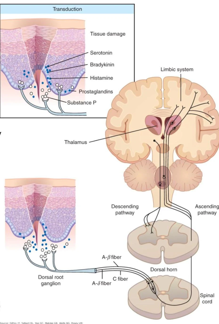
Second-order neurons: Located in the dorsal horn of the spinal cord. These neurons decussate (cross over to the opposite side) and ascend via the STT to the thalamus.
Third-order neurons: Project from the thalamus to the somatosensory cortex.
This diagram illustrates the process of pain transduction, showing how tissue damage leads to the release of neurotransmitters like serotonin, bradykinin, histamine, prostaglandins, and substance P. These signals are transmitted via nerve fibers (A-beta, A-delta, and C fibers) through the dorsal root ganglion and spinal cord to the thalamus and limbic system in the brain.
4. Perception
Perception is the final stage where the brain interprets the signal. This occurs in the somatosensory cortex, which provides the sensory dimension (location and intensity), and the limbic system, which provides the affective (emotional) dimension, such as fear or suffering.
5. Modulation
The body does not passively receive pain signals; it actively attempts to dampen them through descending modulation pathways. These pathways originate in the brain and descend to the dorsal horn of the spinal cord to inhibit nociceptive neurons.
Key inhibitory regions and their neurotransmitters include:
Locus Coeruleus: Utilizes epinephrine (norepinephrine).
Exam Tip: Remember that modulation can be both inhibitory (reducing pain) or excitatory (amplifying pain). The descending pathways mentioned above are primarily inhibitory mechanisms that act via direct inhibition or through local interneurons in the dorsal horn.
Nerve Injury and Abnormal Pain Responses
While acute pain is typically protective, nerve injury (such as from diabetes, shingles, or cancer) can lead to pathological states known as central sensitization. In this state, continuous afferent signaling causes the spinal cord to become hyper-excitable, resulting in a higher output to the brain for even minor inputs.
This leads to two distinct clinical phenomena:
Hyperalgesia: An exaggerated, increased pain intensity in response to a stimulus that is normally painful.
Allodynia: A painful response to a stimulus that is normally innocuous (e.g., a light touch or the brush of clothing).
Diagram illustrating the process of nerve injury leading to spontaneous dysesthesias and allodynia through peripheral nerve degeneration and spinal sensitization.
Clinical Correlation: Referred Pain
A fascinating byproduct of the spinal architecture is referred pain. Because cutaneous (skin) and visceral (organ) sensory neurons often converge onto the same second-order projection neuron in the spinal cord, the brain may misinterpret the source of the signal. Consequently, a person experiencing a heart attack may feel pain in their left arm rather than their chest.
Diagrams illustrating referred pain patterns in the human torso, showing areas of pain associated with internal organs.
Keep reading for free.
Create an account to finish this reading and make it part of your study routine.