Deep Vein Thrombosis and Pulmonary Embolism: Clinical Management and Nursing Care
This content explains the pathophysiology, risk factors, clinical manifestations, diagnostic studies, and nursing management of Deep Vein Thrombosis (DVT) and Pulmonary Embolism (PE). It covers anticoagulation, antiplatelet therapy, and patient education for preventing and managing these conditions.
Hematology involves the complex processes of hematopoiesis (the production of blood cells) and hemostasis (the body's mechanism for stopping bleeding through clot formation). While clotting is a vital physiological response to injury, abnormal clotting within the vascular system can lead to life-threatening complications. Understanding the distinction between venous and arterial thrombosis is fundamental to providing safe and effective nursing care for clients at risk of thromboembolic events.
Understanding Thrombosis and Embolism
A thrombus is a blood clot that develops in a location that can block necessary blood flow. When this occurs in an artery, it is termed an arterial thrombosis; when it occurs in a vein, it is known as a venous thromboembolism (VTE). VTE encompasses two primary conditions: Deep Vein Thrombosis (DVT) and Pulmonary Embolism (PE).
Deep Vein Thrombosis (DVT) occurs when a clot forms in a large vein, most commonly in the lower extremities. DVTs can be classified as:
Proximal: Occurring above the knee (e.g., femoral or iliofemoral veins), which carries a higher risk of migrating to the lungs.
Distal: Occurring below the knee, which carries a lower risk of migration.
Provoked: Caused by an acquired factor like surgery or obesity.
Unprovoked: Occurring without an identifiable risk factor.
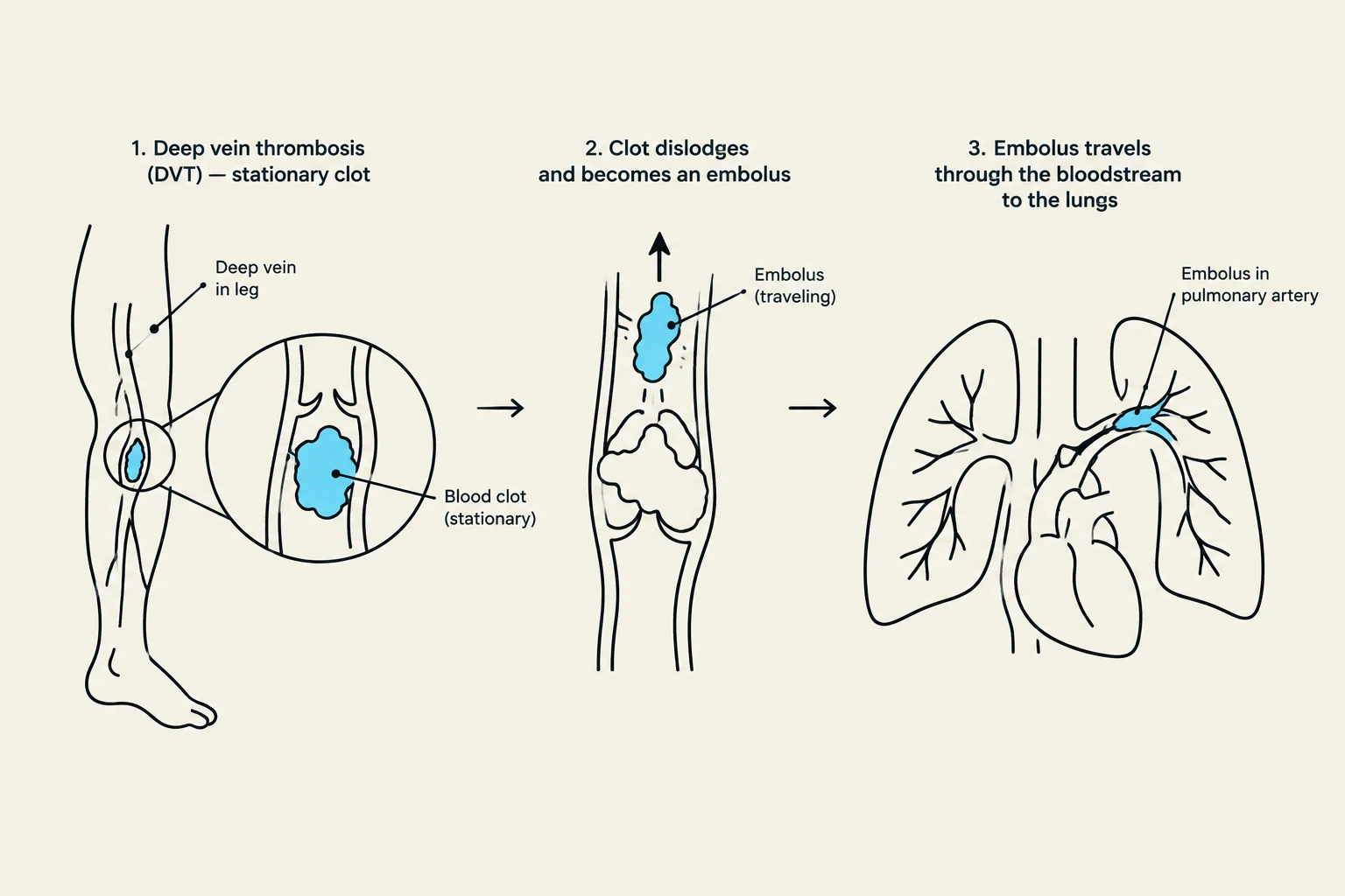
If a thrombus dislodges and travels through the bloodstream, it becomes an embolism. If that embolus travels to the lungs, it results in a Pulmonary Embolism (PE), a medical emergency.
Diagram illustrating the process of deep vein thrombosis (DVT) leading to a pulmonary embolism.
Risk Factors and Comorbidities
The risk of developing a blood clot is influenced by several factors that either increase blood viscosity, decrease blood flow, or trigger hypercoagulability.
Individual and Environmental Factors
Immobility: Prolonged bed rest, surgery, or long airplane flights reduce blood flow and increase risk.
Lifestyle: Smoking and obesity (which increases pressure on lower extremities) are significant contributors.
Genetics: Mutations such as Factor V Leiden (FVL) cause the blood to clot abnormally.
Comorbidities
Certain medical conditions increase the likelihood of thrombosis:
Malignancy: Cancers of the lung, pancreas, or blood (lymphoma/myeloma).
Cardiovascular/Respiratory: Heart failure, atrial fibrillation (A-fib), and obstructive airway disease.
Other: Diabetes mellitus, kidney disease, and systemic autoimmune disorders like systemic sclerosis (scleroderma).
Nursing Tip: Always assess for infection. The body's inflammatory response to infections (such as pneumonia or UTIs) can trigger platelet activation and fibrin deposits, leading to thrombus formation.
Clinical Manifestations and Complications
The location of the clot determines the clinical presentation. Nurses must be able to distinguish between different life-threatening events to prioritize care.
Condition
Primary Clinical Manifestations
DVT
Unilateral leg swelling, pain, redness, and warmth
PE
Sudden shortness of breath, chest pain (worsens with deep breathing), tachycardia, and hemoptysis
MI (Heart Attack)
Chest pain radiating to the left arm, shortness of breath, and diaphoresis
Ischemic Stroke
Facial weakness, difficulty speaking, visual deficits, and seizures
If a client experiences a severe hemorrhage (losing approximately one-fifth of their blood volume), they may enter hypovolemic shock, a state where the heart cannot pump enough blood to sustain organ function. Furthermore, a severe systemic condition called Disseminated Intravascular Coagulation (DIC) can occur, where clotting proteins become overactive, leading to uncontrolled bleeding.
This illustration depicts the relationship between Deep Vein Thrombosis (DVT) and Pulmonary Embolism (PE). DVT occurs when a blood clot forms in a deep vein, often in the leg, leading to symptoms like swelling and warmth. This clot can travel through the bloodstream to the lungs, causing a Pulmonary Embolism, which blocks a pulmonary artery and can result in shortness of breath and chest pain.
Diagnostic Studies
When a clinician suspects a thromboembolic event, several diagnostic tools are utilized:
D-dimer Test: A blood test that detects a protein fragment produced when a clot dissolves. An elevated result suggests a clot is present, though it can also be elevated due to pregnancy or recent surgery.
Venous Doppler Ultrasound: Used to provide a clear view of the veins and blood flow to identify a DVT.
Computed Tomography (CT) Scan: Provides a detailed, often 3-D (spiral CT) view of the body to identify clots in the lungs or brain.
Magnetic Resonance Angiography (MRA): A noninvasive imaging test that specifically examines the blood vessels.
V/Q Scan (Ventilation/Perfusion): A test using small amounts of radioactive substances to check how well air and blood are moving through the lungs.
Pharmacological Management
Treatment focuses on preventing the growth of existing clots and the formation of new ones.
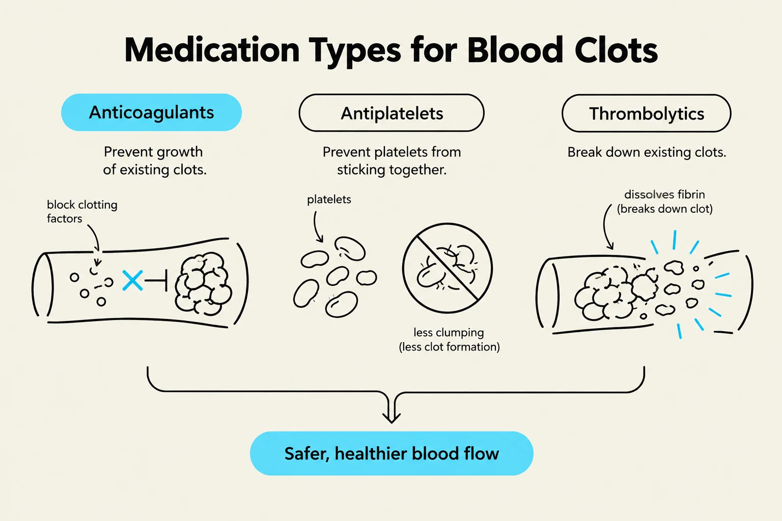
Anticoagulants
Warfarin (Coumadin): An oral anticoagulant that requires frequent monitoring of the International Normalized Ratio (INR). A therapeutic INR is typically between 2 and 3 (or 2.5 to 3.5 for clients with mechanical heart valves). The antidote for warfarin is Vitamin K.
Direct Oral Anticoagulants (DOACs): Medications like apixaban and rivaroxaban are often preferred because they do not require routine INR monitoring or strict dietary changes. However, they are not used in pregnancy or severe kidney disease.
Antiplatelets and Thrombolytics
Antiplatelet Medications: Drugs like aspirin and clopidogrel (Plavix) prevent platelets from sticking together.
Thrombolytics: These are "clot-busting" medications used to dissolve clots that have already formed.
This diagram illustrates three types of medications for blood clots: anticoagulants, antiplatelets, and thrombolytics, and their mechanisms of action to promote safer, healthier blood flow.
Nursing Management and Patient Education
Nursing Interventions
Prevention: For postoperative or immobile clients, use Sequential Compression Devices (SCDs) and encourage early ambulation to maintain peripheral circulation.
Assessment: Prioritize the ABCs (Airway, Breathing, Circulation). Monitor oxygen saturation (goal ≥95%), vital signs (tachycardia/tachypnea), and neurological status.
Safety: When administering anticoagulants, monitor for signs of bleeding, such as black/tarry stools, abdominal pain, or sudden vision changes.
Patient Education
Education is critical to prevent recurrence and manage medication side effects:
Dietary Consistency: For clients on warfarin, they should maintain a consistent intake of Vitamin K (found in green leafy vegetables like spinach and broccoli) rather than making sudden, large changes.
Lifestyle: Encourage smoking cessation, hydration, and an active lifestyle.
Warning Signs: Teach clients to report any unusual bruising, bleeding gums, or signs of a new clot immediately.
Exam Alert: If a client on warfarin presents with an INR of 4.9 or higher, this is a critical value indicating a high risk for life-threatening hemorrhage. The nurse should be prepared to administer the reversal agent, Vitamin K, as prescribed.
Keep reading for free.
Create an account to finish this reading and make it part of your study routine.