Explores the structure, function, and classification of glandular epithelia, including exocrine and endocrine glands, their modes of secretion, and their roles in cell communication and homeostasis.
Glandular epithelia are specialized tissues designed for a single primary purpose: secretion. These cells synthesize and release various products—ranging from proteins and mucins to steroid hormones—that act on surfaces, within internal lumens, or on distant target tissues to facilitate cell communication and maintain homeostasis. Understanding the distinction between exocrine and endocrine systems is fundamental to studying how these tissues maintain the body's internal environment.
Classification of Glandular Epithelia
Glandular epithelia are broadly categorized based on their method of secretion and whether they utilize a duct system.
Exocrine Glands
Exocrine glands release their products onto an epithelial surface or into an organ lumen. This secretion is typically delivered via a duct system. Exocrine glands can be classified by several criteria:
Number of cells: They may be unicellular (such as the Goblet cell, which releases mucus directly onto a surface) or multicellular.
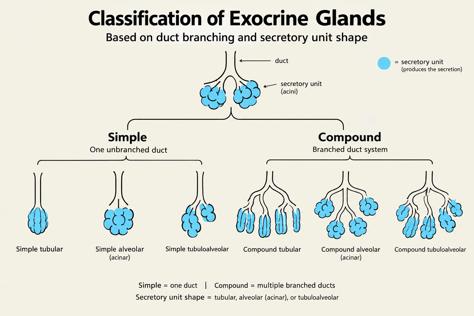
Duct structure: They can be simple (unbranched) or compound (branched).
Secretory unit shape: The shape of the secretory portion can be tubular, acinar, or alveolar.
Mode of secretion: This refers to how the cell releases the product:
Merocrine: Release via exocytosis.
Apocrine: Release involves the apical portion of the cell.
Holocrine: The entire cell breaks down to release its contents.
This diagram illustrates the classification of exocrine glands based on their duct branching and secretory unit shape, categorizing them into simple and compound types with various structural subtypes.
Endocrine Glands
In contrast, endocrine glands are ductless. Instead of traveling through a tube, their secretory products (hormones) are released into the interstitial fluid, where they then enter the bloodstream to reach distant target tissues. Endocrine glands can be organized as small clusters of cells, organized endocrine organs, or even scattered cells within other epithelia, known as the Diffuse Neuroendocrine System (DNES).
Cell Communication and Signaling Modes
The way a secreted molecule reaches its target defines the scale of the communication. There are three primary modes of signaling:
Endocrine: Hormones are transported through the bloodstream to act on distant sites.
Paracrine: Signals are released by cells to affect neighboring cell types locally, without entering the general circulation.
Autocrine: The target of the signal is the cell that secreted it.
Exam Tip: When distinguishing between endocrine and paracrine signaling, look at the transport mechanism. If the signal requires the circulatory system to reach its target, it is endocrine; if it acts on immediate neighbors via interstitial fluid, it is paracrine.
Hormones and Their Mechanisms of Action
Hormones are chemically diverse, and their structure dictates how they interact with target cells.
Chemical Classes of Hormones
Peptides and Proteins: Chains of amino acids (e.g., Insulin, Growth Hormone, ACTH). These are usually stored in granules and released via exocytosis.
Steroids: Derived from cholesterol (e.g., Cortisol, Testosterone, Estrogen). They are generally synthesized on demand.
Amino Acid Derivatives: Modified amino acids, such as catecholamines (epinephrine) and thyroid hormones (T3, T4).
Receptor Interaction
The chemical nature of the hormone determines its receptor location:
Membrane Receptors: Peptide hormones and catecholamines are water-soluble and cannot cross the plasma membrane. They bind to receptors on the cell surface, often triggering a second messenger system (like cyclic AMP) to initiate a cellular response.
Intracellular Receptors: Steroid and thyroid hormones are lipid-soluble and can enter the cell. They bind to receptors in the cytoplasm or nucleus, directly influencing gene transcription and protein synthesis.
This diagram illustrates two distinct hormone signaling pathways: one involving peptide hormones that bind to cell membrane receptors and initiate intracellular cascades, and another involving steroid hormones that diffuse into the cell, bind to intracellular receptors, and directly influence gene expression.
Homeostasis and Regulation
Glandular epithelia are central to maintaining homeostasis—the stable internal environment required for life (e.g., body temperature, blood pressure, and fluid volume). This regulation involves a feedback loop consisting of a receptor (sensor), a control center (integrator), and an effector (responder).
Regulation can be:
Intrinsic: Autoregulation where cells adapt automatically to environmental changes.
Extrinsic: Mediated by the nervous system (rapid, short-term) or the endocrine system (slower, long-lasting).
Key Endocrine Organs and Histology
The Hypothalamic-Pituitary Axis
The hypothalamus and pituitary gland form a critical regulatory axis.
Anterior Pituitary (Adenohypophysis): Composed of glandular epithelial tissue arranged in cords or clumps. It is regulated by the hypothalamus via the hypophyseal portal system, a specialized microcirculation that delivers releasing hormones directly to the anterior lobe.
Posterior Pituitary (Neurohypophysis): This is an extension of the central nervous system. It does not synthesize hormones; instead, it stores and releases hormones (ADH and Oxytocin) produced by hypothalamic neurons.
The Thyroid Gland
The thyroid is a bilobate gland characterized by unique follicles. These follicles are filled with colloid, an extracellular storage form of thyroglobulin.
Follicular cells produce T3 and T4, which increase metabolism.
Parafollicular cells (C cells) produce calcitonin, which lowers blood calcium levels.
The Pancreas
The pancreas serves both exocrine and endocrine functions. The endocrine portion consists of the Islets of Langerhans.
Alpha (α) cells: Produce glucagon to increase blood glucose.
Beta (β) cells: Produce insulin to decrease blood glucose.
Delta (δ) cells: Produce somatostatin, which exerts a paracrine effect on α and β cells.
The Adrenal Glands
Located atop the kidneys, these glands consist of two distinct regions:
Adrenal Cortex: Produces steroid hormones in three layers: the zona glomerulosa (aldosterone), zona fasciculata (cortisol), and zona reticularis (androgens).
Adrenal Medulla: Contains chromaffin cells that release catecholamines (epinephrine and norepinephrine) to mediate the "fight-or-flight" response.
Keep reading for free.
Create an account to finish this reading and make it part of your study routine.