Cytokines and the Mechanisms of the Inflammatory Response
Explore the inflammatory response as a cornerstone of innate immunity, detailing its purpose, cardinal signs, and the crucial role of cytokines as chemical messengers. Understand the coordination between innate and adaptive immunity, including cell-mediated and antibody-mediated responses, and the mechanics of T cell a
When your body experiences a cellular injury—whether from a physical cut, a viral infection, or a bacterial invasion—it does not remain passive. Instead, it launches a sophisticated, multi-layered defense system designed to contain the damage and initiate repair. Central to this defense is the inflammatory response, a rapid, non-specific reaction that serves as a cornerstone of our innate immunity.
The Inflammatory Response: Purpose and Signs
The inflammatory response is a fundamental physiological reaction to any form of cellular injury. Rather than being a random side effect of injury, inflammation is a highly organized process with three primary functional objectives:
Isolation: It works to isolate the damaged area to prevent the spread of pathogens or toxins.
Cleanup: It facilitates the removal of damaged or dead cells.
Repair: It recruits the necessary cells and proteins to the site to begin the process of tissue repair.
While inflammation is essential for healing, it is often uncomfortable. Clinicians identify the presence of inflammation through four cardinal signs:
Redness: Caused by increased blood flow to the area.
Heat: A result of increased metabolic activity and blood flow.
Swelling: Occurs as fluids and proteins leak into the tissue.
Pain: Triggered by chemical mediators and pressure on nerve endings.
Study Tip: Remember that inflammation is a component of innate (nonspecific) immunity. It responds to pathogens the same way regardless of whether it has encountered them before, and it is typically the dominant response during the first 12 hours of exposure.
Cytokines: The Chemical Messengers
If the inflammatory response is the "army" moving to a battlefield, cytokines are the "radio signals" coordinating the movement. Cytokines are proteins produced by immune cells that enhance and regulate immune responses.
These signaling molecules are responsible for many of the systemic symptoms we associate with being sick. When cytokines enter the bloodstream, they can induce "flu-like" symptoms, including aches and chills. One of the most significant systemic effects is fever. Fever is considered an innate response to cellular injury, triggered by substances called pyrogens. Pyrogens are released from damaged cells or bacteria and act on the hypothalamus in the brain to set a higher body temperature.
There are several key types of cytokines that play distinct roles in the immune landscape:
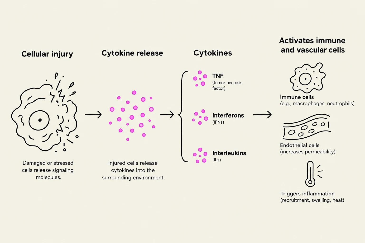
Tumor Necrosis Factor (TNF): A potent signaling protein involved in systemic inflammation.
Interferons: Critical for antiviral responses.
Interleukins: A large group of cytokines that facilitate communication between different white blood cells.
This diagram illustrates the process of cellular injury leading to cytokine release, which then activates immune and vascular cells, ultimately triggering inflammation.
Coordinating the Defense: Innate vs. Adaptive Immunity
The body’s ability to defend itself relies on the coordination between two distinct but overlapping systems: Innate Immunity and Adaptive Immunity.
Innate Immunity
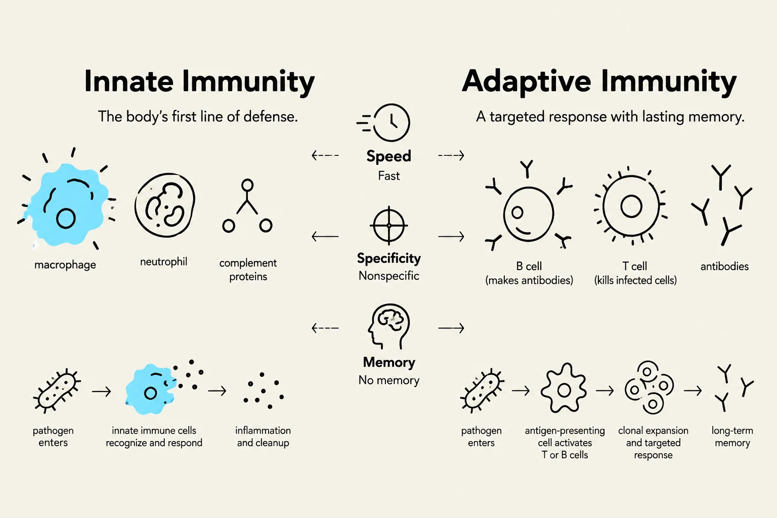
Innate immunity is "nonspecific," meaning it responds to all pathogens in a similar manner. It is incredibly fast, responding within seconds to hours. This system utilizes various cells, such as phagocytes (macrophages, neutrophils, eosinophils, and dendritic cells) and non-phagocytic cells (Natural Killer cells, basophils, and mast cells).
Adaptive Immunity
Unlike the innate response, adaptive immunity (also known as acquired immunity) is highly specific. It takes longer to mobilize—usually 3 to 5 days after exposure to an antigen—but it possesses the remarkable capacity for immunological memory. Adaptive immunity is divided into two specialized mechanisms:
Cell-Mediated Immunity: This involves the action of T cells. These cells are responsible for responding to intracellular threats like viruses, intracellular bacteria, cancer cells, and foreign transplanted organs.
Antibody-Mediated Immunity: This involves B cells and the production of antibodies to target pathogens in the extracellular spaces.
This diagram illustrates the differences between innate and adaptive immunity, detailing their respective components, speed, specificity, and memory capabilities.
Deep Dive: The Mechanics of Cell-Mediated Immunity
To understand how the body targets specific threats, we must look at how T cells are activated and how they function. T cells mature in the thymus gland after migrating from the bone marrow. During this maturation, the body ensures immunocompetence (the ability to recognize antigens) and self-tolerance (the destruction of T cells that would attack the body's own cells).
T cells recognize antigens through docking sites called Major Histocompatibility Complex (MHC) glycoproteins. There are two critical classes of MHC:
Class I MHC: Found on nearly all nucleated cells. These present antigens from inside the cell (endogenous) to Cytotoxic T (TC) cells.
Class II MHC: Found only on antigen-presenting cells (like dendritic cells). These present antigens taken in from outside the cell (exogenous) to Helper T (TH) cells.
The Roles of Helper and Cytotoxic T Cells
The coordination of the entire immune response often rests on the Helper T (TH) cell. TH cells do not kill pathogens directly; instead, they secrete cytokines (such as various interleukins) that activate and enhance both innate immunity and the adaptive responses of B cells and T cells.
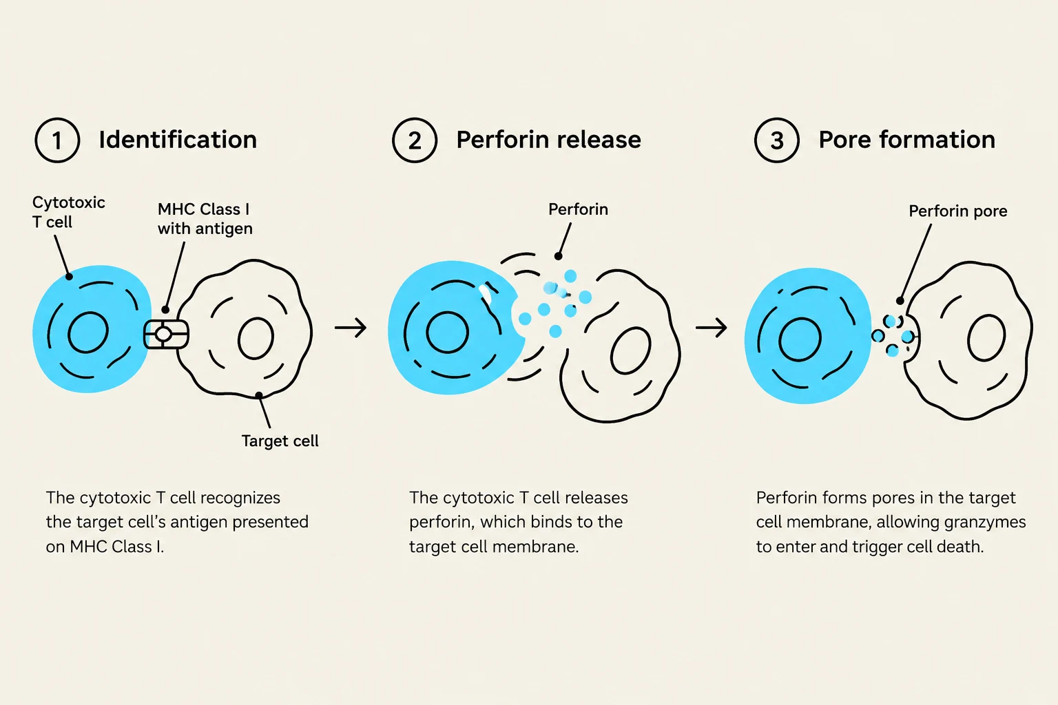
In contrast, Cytotoxic T (TC) cells are the "assassins" of the immune system. When a TC cell recognizes a diseased cell presenting a foreign antigen on a Class I MHC molecule, it executes a precise four-step process:
Binding: The TC cell binds to the target cell.
Pore Formation: It releases perforin, which creates holes in the target cell's plasma membrane.
Degradation: It releases enzymes that enter the target cell to fragment its DNA and degrade proteins.
Detachment: The target cell begins to degrade, and the TC cell detaches to search for its next target.
This diagram illustrates the process of cytotoxic T cell-mediated cell killing, involving identification of the target cell via MHC Class I, release of perforin, and pore formation in the target cell membrane to allow granzyme entry and cell death.
Clinical Connection: This mechanism explains why organ transplantation is so complex. In allografts (transplants between non-identical individuals of the same species), the recipient's TC cells will recognize the donor's MHC molecules as foreign antigens, leading to organ rejection and necrosis unless immunosuppressive therapy is used.
Keep reading for free.
Create an account to finish this reading and make it part of your study routine.