Coronary Artery Disease: Pathophysiology, Assessment, and Interventions
This comprehensive guide explains Coronary Artery Disease (CAD), its risk factors, and prevention strategies. It details myocardial infarction assessment, diagnostic markers, and various surgical and interventional options like CABG and TMR. The content also covers post-operative care, potential complications, and esse
Coronary Artery Disease (CAD) is the most common type of cardiovascular disease in the United States and serves as a major cause of death. It is a progressive condition characterized by the deposition of lipids on the intima of major arteries, which triggers an inflammatory response. As this process continues, a fibrous cap develops over a fatty core, forming a plaque that can protrude into the arterial lumen. If this plaque ruptures, it can lead to a thrombus, myocardial infarction (MI), or sudden death.
This diagram illustrates the progression of coronary artery plaque formation and rupture, showing lipid accumulation in the intima, the development of a mature plaque with a fibrous cap, and subsequent plaque rupture leading to thrombus formation.
Risk Factors and Prevention
Understanding the risk factors for CAD is essential for both prevention and management. Risk factors can be categorized by how they affect the heart's workload and oxygen demand.
Modifiable Risk Factors
Several lifestyle-related factors can be managed to curtail the progression of CAD:
Hypertension: Increases the workload of the heart.
Cigarette Smoking: A significant contributor to vascular damage.
Obesity: Increases myocardial oxygen demand.
Elevated Homocysteine Levels: Linked to increased risk.
Diet Therapy
Dietary modification is a cornerstone of management. Patients should be taught to follow a diet that is:
Low in fat and cholesterol.
Low in sodium.
Rich in healthy fats: Patients should prioritize fats from sources like olive oil or nuts.
Exam Tip: When evaluating patient teaching regarding diet, look for the answer that emphasizes healthy fat sources (like olive oil/nuts) rather than simply eliminating all fats or red meats.
Myocardial Infarction (MI) Assessment
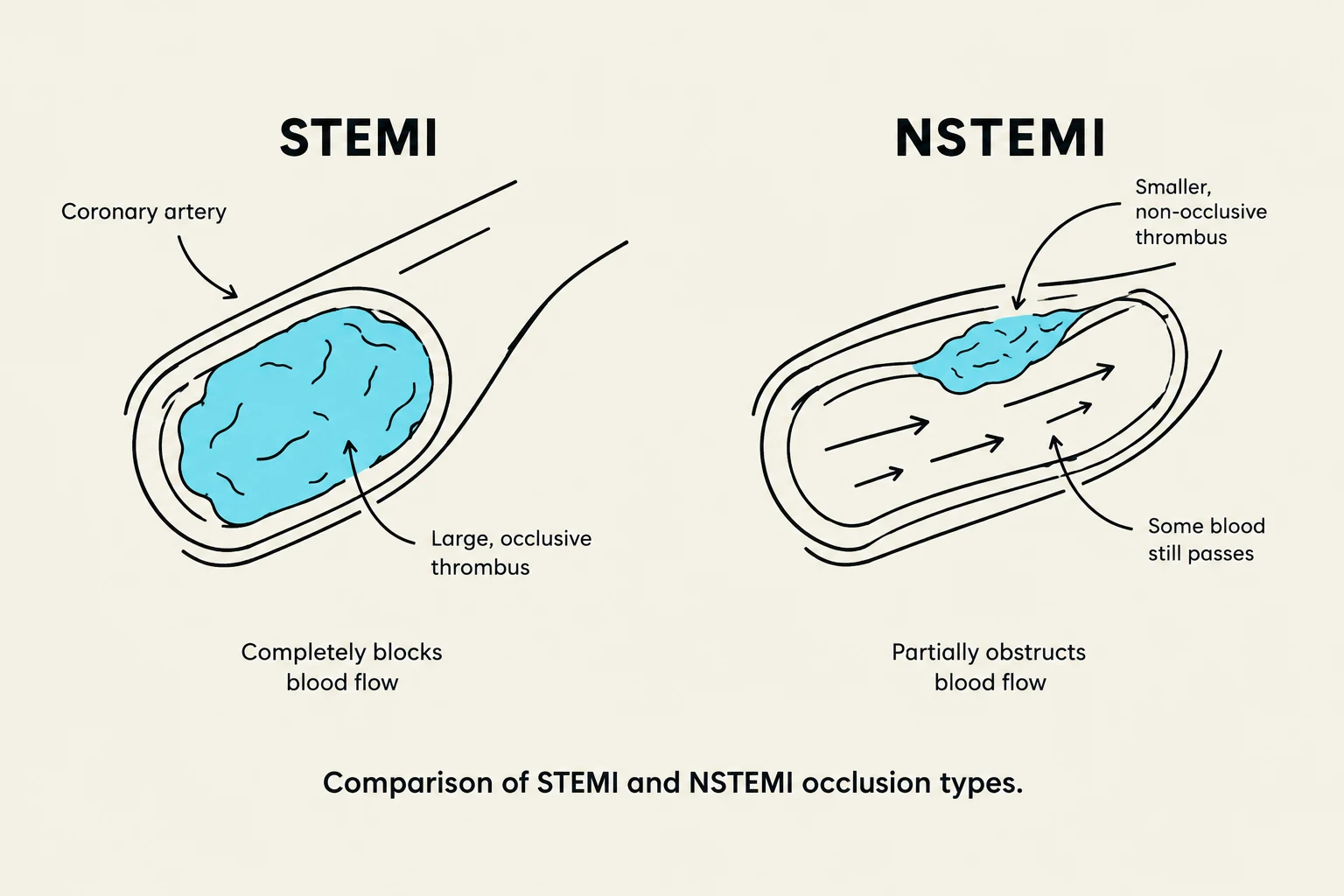
Comparison of STEMI and NSTEMI occlusion types.
A myocardial infarction occurs when there is a sustained interruption of blood flow, leading to irreversible myocardial cell death (necrosis). This can be classified as a STEMI (ST-segment elevation MI), caused by an occlusive thrombus, or an NSTEMI (non-ST-segment elevation MI), caused by a non-occlusive thrombus.
Physical Assessment
When assessing a patient for a potential MI, nurses should look for a variety of clinical manifestations:
Pain: Often substernal or epigastric.
Respiratory/Circulatory: Shortness of breath, pallor, diaphoresis, and pulse changes/dysrhythmias.
Gastrointestinal: Nausea and vomiting.
Neurological/Psychological: Dizziness, lightheadedness, weakness, and a "feeling of doom."
Diagnostic Markers
To confirm an MI, clinicians rely on serial laboratory tests and EKG changes:
Troponin: The most reliable indicator; it elevates quickly and stays elevated the longest.
Creatine Kinase (CK-MB): Specifically indicates heart muscle damage; an MB index >4%−6% of total CK is indicative of a cardiac event.
Myoglobin: An early marker that elevates within 2 hours of injury.
12-Lead EKG: Used to determine the location and size of the infarction through ST-segment elevation, T-wave inversion, or Q waves.
Surgical and Interventional Options
When medical management and Percutaneous Coronary Intervention (PCI)—such as balloon angioplasty or stenting—are insufficient, surgical revascularization may be necessary.
Coronary Artery Bypass Graft (CABG)
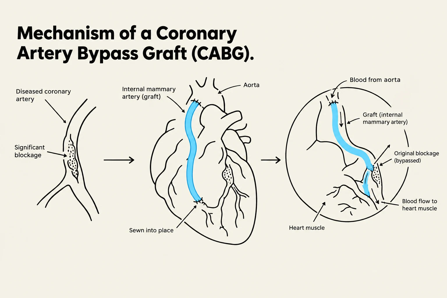
Diagram illustrating the mechanism of a Coronary Artery Bypass Graft (CABG) procedure, showing a diseased coronary artery with a blockage, the placement of an internal mammary artery graft, and the resulting bypass of the blockage to restore blood flow to the heart muscle.
CABG uses a healthy vessel (such as the internal mammary artery, saphenous vein, or radial artery) to bypass a diseased section of the coronary artery.
Traditional CABG: Requires a sternotomy and the use of a cardiopulmonary bypass (CPB) machine to stop the heart during surgery.
Off-Pump Coronary Artery Bypass (OPCAB): Performed while the heart continues to beat, avoiding the use of a heart-lung machine. This requires specialized tools, including a heart stabilizing device and a sternal retractor.
Minimally Invasive Direct CABG (MIDCAB): Does not involve a sternotomy or CPB and is typically used for disease in the left anterior descending or right coronary artery.
Robotic/Totally Endoscopic (TECAB): Minimally invasive approaches to revascularization.
Transmyocardial Laser Revascularization (TMR)
TMR is an indirect revascularization method where a high-energy laser is used to burn channels into the heart muscle (but not through it) to facilitate blood flow to ischemic areas.
Post-Operative Care and Complications
Complications
Patients are at risk for several serious complications following an MI or cardiac surgery:
Dysrhythmias: Atrial fibrillation is particularly common after CABG.
Hemodynamic Issues: Heart failure, cardiogenic shock, and perfusion deficits to major organs.
Post-Surgical Specifics: Bleeding, infection, pulmonary complications, and Dressler Syndrome.
Cognitive Changes: Patients may experience impaired memory or a short attention span for 6–8 weeks post-operation.
Nursing Interventions
Post-operative care in the ICU involves intensive monitoring, including:
Hemodynamics: Continuous ECG, pulmonary artery catheters (Swan-Ganz), and arterial lines.
Respiratory Support: Endotracheal tubes for mechanical ventilation and monitoring SpO2.
Drainage: Monitoring pleural and mediastinal chest tubes for bleeding or wound healing.
Cardiac Rehabilitation and Patient Teaching
Successful recovery depends on comprehensive cardiac rehabilitation and patient education.
Physical Activity
Nurses should guide patients in resuming activity using the METs scale. It is vital that patients understand the difference between:
Isotonic activities: Involving muscle contraction and movement.
Isometric activities: Involving muscle contraction without movement.
Discharge Teaching
Patients and caregivers should be educated on the following:
Wound Care: Monitoring sternal and leg incisions for redness, pus, or fever.
Medication Adherence: Ensuring compliance with prescribed regimens.
Pain Management: Balancing comfort with the need for respiratory hygiene.
Lifestyle Resumption: Gradual return to daily activities and sexual activity.
Evaluating Success
The effectiveness of the intervention and rehabilitation is measured by:
Stable vital signs and EKG returning to baseline.
Resolution of pain and decreased anxiety.
The patient's ability to effectively manage their therapeutic regimen and return to a functional quality of life.
Keep reading for free.
Create an account to finish this reading and make it part of your study routine.