Coronary Artery Disease and Angina: Understanding Ischemia and Clinical Presentation
This content explains Coronary Artery Disease (CAD), its pathophysiology involving atherosclerosis and ischemia, and the clinical manifestations of angina and angina equivalents. It also details the classification of angina (chronic stable, unstable, variant) and Acute Coronary Syndrome (ACS), differentiating between N
Coronary Artery Disease (CAD) is the leading cause of death in the United States, accounting for one in four deaths. At its core, CAD is a progressive condition characterized by the narrowing or occlusion of the coronary arteries, typically due to the buildup of atherosclerotic plaques. This narrowing creates a critical imbalance: the heart muscle's demand for oxygen exceeds the supply provided by the blood flow. When this mismatch occurs, the patient experiences ischemia, the clinical precursor to more severe cardiac events.
The Pathophysiology of Ischemia and Atherosclerosis
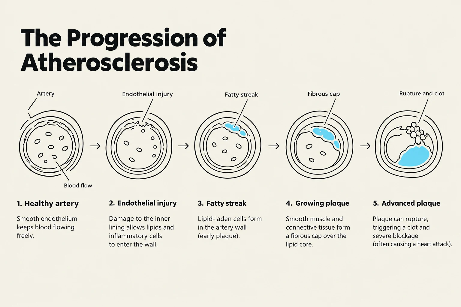
Atherosclerosis begins with injury to the endothelium, the inner lining of the vessel wall. This injury can be caused by smoking, high cholesterol, hypertension, or diabetes. Over time, lipids (specifically LDL cholesterol) are transported into the arterial intima, forming a fatty streak. As the process progresses, collagen covers these lipids to form a fibrous plaque.
If a plaque becomes unstable, it can rupture, leading to the formation of a thrombus (clot) that can cause total occlusion of the artery. The severity of the blockage dictates the clinical outcome:
Stenosis: A blockage of $70%$ or more in a standard artery typically causes ischemia.
Left Main Coronary Artery: Because this artery supplies a massive portion of the heart, a blockage of even $50%$ or more can trigger significant angina.
When blood flow is insufficient, the myocardium enters a state of hypoxia (oxygen deprivation). Within just 10 seconds of total occlusion, the heart cells shift from aerobic metabolism (using oxygen) to anaerobic metabolism. This metabolic shift results in the accumulation of lactic acid, which contributes to the sensation of pain. If blood flow is not restored within approximately 20 minutes, irreversible heart muscle damage begins, provided there is insufficient collateral circulation.
Diagram illustrating the progression of atherosclerosis, from a healthy artery to an advanced plaque with rupture and clot.
Clinical Manifestations: Angina and Angina Equivalents
Angina is the clinical manifestation of myocardial ischemia, characterized by severe chest pain. Patients rarely describe it as "sharp"; instead, they typically report sensations of pressure, heaviness, squeezing, or tightness. While the pain is most commonly substernal (behind the breastbone), it frequently radiates to the jaw, neck, shoulders, arms, or the interscapular region (between the shoulder blades).
It is vital for clinicians to recognize angina equivalents. Not all patients present with classic chest pain. Women and older adults, in particular, may present with atypical symptoms such as:
Dyspnea (shortness of breath)
Nausea
Extreme fatigue
Exam Tip: If a patient describes "indigestion" or "burning" in the epigastric region along with back pain between the shoulder blades, do not dismiss it as gastrointestinal. These are common atypical presentations of cardiac ischemia.
Classifying Angina and Acute Coronary Syndrome (ACS)
The clinical presentation of ischemia is categorized based on the pattern of pain and the underlying degree of arterial blockage.
Chronic Stable Angina
This is predictable pain that occurs during physical exertion, emotional stress, or intense activity. The pain is intermittent, lasts only a few minutes, and is relieved by rest or the administration of sublingual nitroglycerin. On an EKG, this may manifest as ST segment depression or T wave inversion.
Unstable Angina and Acute Coronary Syndrome (ACS)
When angina becomes unpredictable, occurs at rest, or increases in frequency, duration, and severity, it is classified as unstable angina. Unstable angina is part of the Acute Coronary Syndrome (ACS) spectrum, a medical emergency that requires urgent intervention.
NSTEMI vs. STEMI
To distinguish between unstable angina and a myocardial infarction (MI), clinicians look at serum cardiac biomarkers (such as troponin) and EKG changes:
Non-ST Segment Elevation MI (NSTEMI): Involves a partial blockage and reduced blood flow. Biomarkers will be elevated, indicating cell death, but the EKG will not show ST elevation.
ST-Elevation MI (STEMI): Indicates a complete, total occlusion of a coronary artery. This is identified by ST elevation on a 12-lead EKG.
Variant (Prinzmetal's) Angina
A rarer form of angina is Prinzmetal's angina, which is caused by sudden coronary artery spasms rather than fixed atherosclerotic plaques. These spasms are often due to increased intracellular calcium and can be triggered by tobacco, alcohol, amphetamines, extreme cold, or even during REM sleep. Unlike stable angina, this occurs at rest and may show transient ST segment elevation on an EKG.
This diagram compares and contrasts four types of angina and myocardial infarction: Chronic Stable Angina, Unstable Angina, NSTEMI, and STEMI. It illustrates the causes, ECG findings, troponin levels, pain characteristics, and typical settings for each condition.
Management Strategies
The primary goal of managing CAD and angina is to balance the myocardial oxygen supply and demand. This is achieved by either increasing oxygen delivery or decreasing the heart's workload.
Pharmacological Interventions
Nitrates: Medications like nitroglycerin are used to cause vasodilation. Sublingual nitroglycerin is used for acute relief of anginal attacks. Note that a common side effect is headache due to cerebral vasodilation.
Beta Blockers: These reduce myocardial oxygen demand by decreasing the heart rate, blood pressure, and contractility.
Calcium Channel Blockers: Used if beta blockers are contraindicated or poorly tolerated, these promote systemic and coronary vasodilation.
Lipid-Lowering Agents: Statins are used to inhibit cholesterol synthesis in the liver, thereby reducing LDL levels.
Summary of Management Goals
The management approach can be summarized by the following relationship:
To prevent ischemia, we must ensure that:
Oxygen Supply≥Oxygen Demand
Clinical Note: When administering sublingual nitroglycerin, if the pain does not subside after the first dose, the patient can take up to three doses, five minutes apart. If the pain persists after three doses, it is a medical emergency requiring immediate hospital evaluation.
Keep reading for free.
Create an account to finish this reading and make it part of your study routine.