An overview of blood composition, including plasma, serum, and cellular components like erythrocytes, leukocytes, and thrombocytes. It also covers their functions and basic histological staining techniques.
Blood is a highly specialized form of connective tissue that circulates throughout the body, serving as a vital lifeline for almost every physiological process. Rather than being a simple liquid, blood is composed of living cellular elements suspended within an extracellular matrix. This complex mixture allows for the efficient transport of life-sustaining materials and the rapid deployment of immune defenses.
The Composition of Blood: Plasma and Serum
The liquid portion of blood is known as plasma, which serves as the extracellular matrix. Plasma is composed of approximately 90% water, with the remaining 10% consisting of dissolved sugars, fats, proteins, hormones, and salts.
One of the most critical roles of plasma is the transport of waste products, such as carbon dioxide (CO2), ammonia, and urea, to organs where they can be recycled or excreted. Plasma also contains essential proteins, most notably fibrinogen, which is a key clotting factor.
Clinical Distinction: Plasma vs. Serum
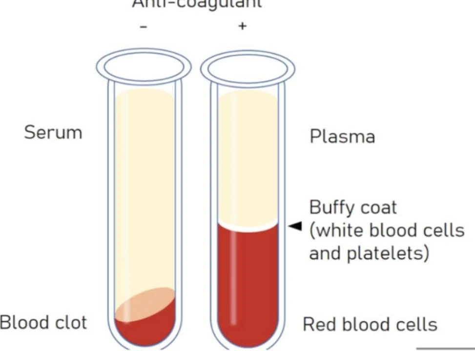
It is important to distinguish between these two terms in a laboratory setting. Plasma contains all clotting factors, including fibrinogen. Serum, however, is defined as plasma that has been stripped of these clotting factors after a blood clot has formed.
This illustration shows the difference between serum and plasma. Serum is the liquid component of blood that remains after clotting, while plasma is the liquid component of blood that remains after an anticoagulant is added, preventing clotting.
The blood's ability to stop bleeding relies on the coagulation cascade, a complex chain reaction of specialized clotting factors that work together to form a stable clot.
Cellular Components of Blood
The cellular portion of blood consists of several distinct types of cells, each specialized for a specific task. These cells are produced through a process called hematopoiesis, which begins in the yolk sac during embryonic development and eventually moves to the bone marrow, where it continues throughout life.
Diagrams of various human blood cells including monocytes, lymphocytes, neutrophils, eosinophils, basophils, macrophages, erythrocytes, and platelets.
Erythrocytes (Red Blood Cells)
Erythrocytes, or RBCs, are the most numerous cells in the blood. They are characterized as small, biconcave discs that lack nuclei and organelles, a structure that maximizes their efficiency. Their cytoplasm is packed with hemoglobin, a protein that allows them to pick up oxygen (O2) at the lungs and release it into body tissues, while simultaneously carrying CO2 back to the lungs.
RBCs have a lifespan of approximately 100–120 days. Once they become worn out, they are removed from circulation by macrophages located in the spleen and liver.
Clinical Note: Blood Disorders
Sickle Cell Anemia: A genetic disorder where a single amino acid substitution (glutamic acid to valine) causes hemoglobin to form long fibers. This distorts the RBCs into rigid, crescent-shaped "sickle cells" that can block blood flow, leading to intense pain and anemia.
Spherocytosis: A condition where RBCs lose their biconcave shape and become spherical due to a defect in the cytoskeleton. These cells are less flexible and often get trapped and destroyed in the spleen, potentially leading to an enlarged spleen (splenomegaly).
Leukocytes (White Blood Cells)
Leukocytes, or white blood cells (WBCs), are the primary agents of the immune system. While they travel through the bloodstream, they often function within the loose connective tissues of organs. Leukocytes are categorized into two main groups based on their appearance:
1. Granulocytes
Granulocytes possess cytoplasmic granules containing enzymes or chemicals, giving them a "grainy" appearance under a microscope. They also typically feature non-spherical, lobed nuclei.
Neutrophils: The most common responders to bacterial invasions, characterized by nuclei with up to five lobes.
Eosinophils: These cells feature bi-lobed nuclei and large granules that stain bright red/pink with acidic dyes. They are vital for fighting parasitic infections and regulating allergic reactions.
Basophils: The rarest leukocyte, these cells contain large, dark purple granules that stain with basic dyes. They release histamine to mediate inflammation during allergic reactions.
2. Agranulocytes
Agranulocytes lack visible cytoplasmic granules, making their cytoplasm appear clear and blue under a microscope.
Lymphocytes: These cells have a large, dark purple nucleus that occupies most of the cell volume. They include B cells (which differentiate into antibody-secreting plasma cells) and T cells (which can directly kill infected cells or activate other immune cells).
Monocytes: The largest of the leukocytes, monocytes have a characteristic C-shaped or kidney-shaped nucleus. When they migrate from the blood into tissues, they differentiate into macrophages (large phagocytic cells) or dendritic cells (professional antigen-presenting cells).
Thrombocytes (Platelets)
Thrombocytes, commonly known as platelets, are not whole cells but rather small, disc-shaped fragments of cytoplasm. They are produced when larger cells called megakaryocytes break apart. Platelets are essential for hemostasis; they plug tears in vessel walls and release granules to recruit more platelets to the site of an injury.
Histological Staining Techniques
To study these components, medical professionals cannot use Gram staining—which is intended for bacteria—because human cells lack the thick peptidoglycan walls required to retain the purple stain. Instead, they use specific histological stains to differentiate cell types based on the acidity or alkalinity of their components:
Wright's Stain and Giemsa Stain: Common routine hematology stains used to visualize blood cells.
Hematoxylin and Eosin (H&E) Stain: A standard histological method.
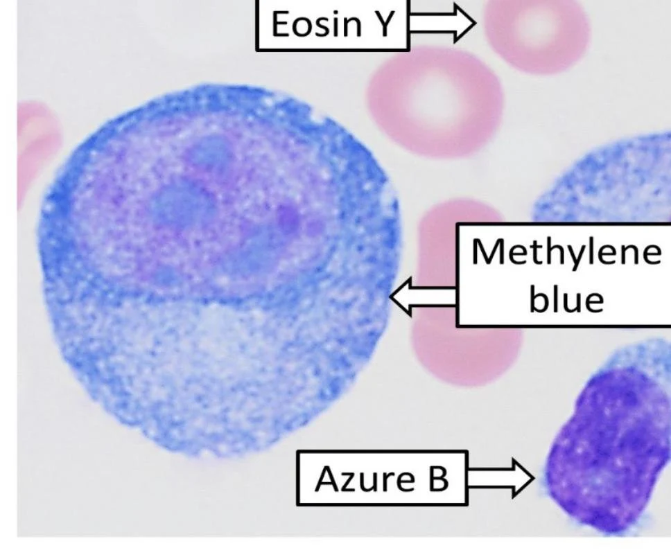
Eosin: An acidic red dye that stains basic components, such as the cytoplasm of erythrocytes and granulocytes.
Methylene Blue: A basic dye that stains acidic structures, such as cell nuclei, blue.
Microscopic view of blood cells stained with Eosin Y, Methylene blue, and Azure B.
Summary of Blood Functions
The multifaceted nature of blood allows it to perform several critical roles simultaneously:
Transport: Moving nutrients, respiratory gases (O2 and CO2), hormones, and immune cells throughout the body.
Waste Removal: Transporting metabolic waste products to organs for excretion or recycling.
Regulation: Helping to maintain and regulate body temperature.
Keep reading for free.
Create an account to finish this reading and make it part of your study routine.