Explains how excitable cells like neurons and muscle fibers use membrane potential, ion gradients, and specialized protein channels (chemical-gated and voltage-gated) to communicate and generate electrical signals such as graded potentials and action potentials.
In the biological world, communication is not just about signals; it is about the movement of electricity. While all living cells maintain a charge difference across their membranes—known as the membrane potential—certain specialized cells are capable of changing this potential deliberately to perform functions like thinking, feeling, or moving. These are known as excitable cells, and they include both neurons (nerve cells) and muscle fibers.
To understand how these cells communicate, we must look at the specialized "gates" that allow ions to flow across the cell membrane, turning chemical and electrical signals into biological action.
The Foundation: Membrane Potential and Gradients
Before a cell can send a signal, it must establish a baseline. This baseline is the resting membrane potential (RMP). In a typical neuron, the inside of the cell is more negative than the outside, often measured at approximately −70 mV.
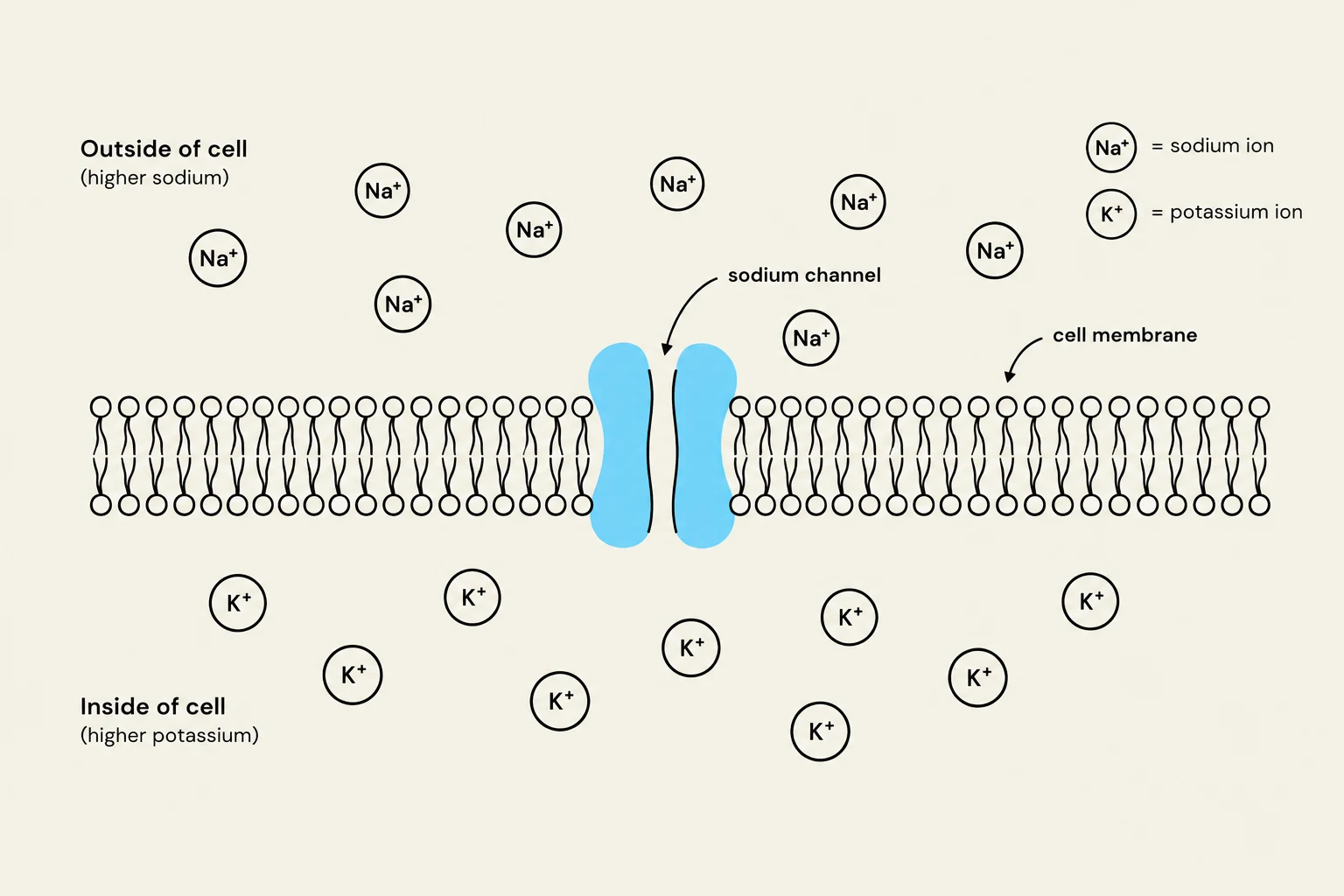
This charge difference exists because of the unequal distribution of ions. While many ions like sodium (Na+), potassium ($K^+$), and calcium (Ca2+) are found on both sides of the membrane, their concentrations differ significantly:
Potassium ($K^+$) is found in much higher concentrations inside the cell.
Sodium (Na+) and other ions are found in much higher concentrations outside the cell.
The movement of these ions is driven by the electrochemical gradient, which is the combination of two forces: the chemical gradient (ions moving from high to low concentration) and the electrical gradient (ions moving toward opposite charges).
Diagram illustrating the cell membrane with sodium and potassium ions, and a sodium channel.
Exam Tip: Remember that the cell membrane is more permeable to potassium than to sodium. This is because there are more potassium "leak" channels present in the membrane, allowing potassium to move more easily than sodium.
Types of Ion Channels: Chemical vs. Voltage Gated
Excitable cells use two primary types of specialized protein channels to manage the flow of ions.
Chemical-Gated Ion Channels
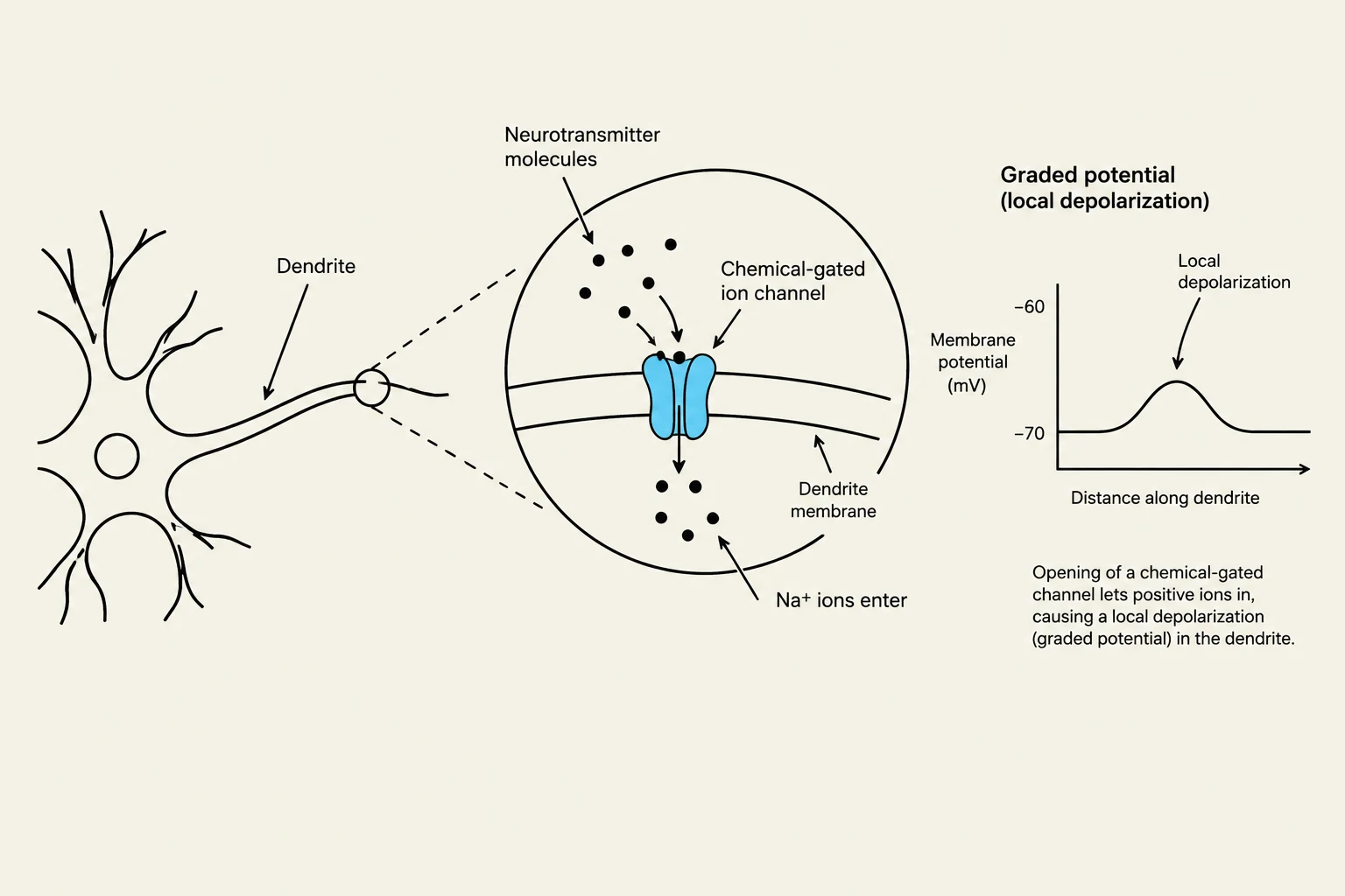
These channels are "triggered" by specific molecules. They remain closed until a specific chemical (such as a neurotransmitter) binds to them, causing the gate to open. In a neuron, these channels are primarily located on the dendrites and the soma (the cell body). In a muscle cell, they are concentrated at the motor end plate.
Voltage-Gated Ion Channels
Unlike their chemical counterparts, these channels respond to changes in the electrical potential of the membrane. They are strategically located along the axon and at the axon terminals of neurons, as well as across most of the membrane of a muscle fiber. These channels are responsible for the rapid propagation of signals over long distances.
Note on the Axon Hillock: There is a specific region of the neuron called the axon hillock that connects the soma to the axon. Interestingly, this region contains neither chemical-gated nor voltage-gated channels.
Graded Potentials: Localized Communication
When a chemical-gated channel opens, ions rush through according to their electrochemical gradients, creating a graded potential. A graded potential is a local electrical change where the strength of the signal depends on how many channels open and how long they stay open.
The direction of ion movement determines the effect on the membrane potential:
Depolarization: If positive ions like sodium (Na+) enter the cell, the inside becomes less negative (more positive). This "raises" the membrane potential toward zero.
Hyperpolarization: If positive ions like potassium ($K^+$) leave the cell, or if negative ions like chloride ($Cl^-$) enter, the inside becomes more negative. This "lowers" the membrane potential further away from zero.
A defining characteristic of graded potentials is that they are local events. The electrical change is greatest at the exact site where the channel opens and diminishes as it moves further away from that site.
This illustration depicts a neuron's dendrite, a magnified view of a chemical-gated ion channel in the dendrite membrane, and a graph showing a local depolarization (graded potential) caused by the influx of positive ions.
Information Flow in Neurons and Muscle Fibers
The way these components work together allows for the efficient transmission of information.
In the Neuron
Information follows a strict directional path:
Dendrites → Soma → Axon → Axon Terminal.
The process begins at the dendrites, where chemical signals trigger graded potentials via chemical-gated channels. If these local changes are strong enough, they travel down the axon via voltage-gated channels, which allow the signal to travel long distances without losing strength. Finally, the signal reaches the axon terminal, where it can trigger the release of chemicals to communicate with the next cell.
In the Muscle Fiber
Muscle cells (or muscle fibers) use a similar logic to initiate movement. The communication occurs at the neuromuscular junction, the site where a neuron meets a muscle cell.
The axon terminal of a neuron binds to the motor end plate of the muscle fiber. This binding opens chemical-gated channels on the motor end plate, creating a graded potential. Because the rest of the muscle fiber is covered in voltage-gated channels, this initial local signal can quickly spread across the entire cell, leading to a muscle contraction.
Diagram illustrating a neuron connecting to a muscle fiber, showing the transmission of a nerve impulse.
Summary Table of Channel Locations
Cell Part
Channel Type
Primary Function
Dendrites / Soma
Chemical-gated
Receive signals and create graded potentials
Axon / Axon Terminal
Voltage-gated
Propagate electrical signals over distances
Motor End Plate
Chemical-gated
Receive signals from the neuron
Muscle Fiber (Body)
Voltage-gated
Spread the signal to trigger contraction
Keep reading for free.
Create an account to finish this reading and make it part of your study routine.