Common Respiratory Disorders and Their Clinical Findings
An overview of common respiratory disorders such as asthma, bronchiolitis, cystic fibrosis, obstructive sleep apnea, and pneumonia, detailing their underlying pathology and key clinical manifestations.
The respiratory system is a complex network of structures designed for efficient gas exchange, ranging from the conductive upper airways to the microscopic alveoli where oxygen enters the bloodstream. When this system is compromised by infection, genetic defects, or airway obstruction, clinicians must rely on a combination of patient history, physical examination findings, and diagnostic imaging to differentiate between various pathologies. This article examines five common respiratory disorders, focusing on their underlying mechanisms and the clinical signs that distinguish them.
Asthma: Variable Airflow Limitation
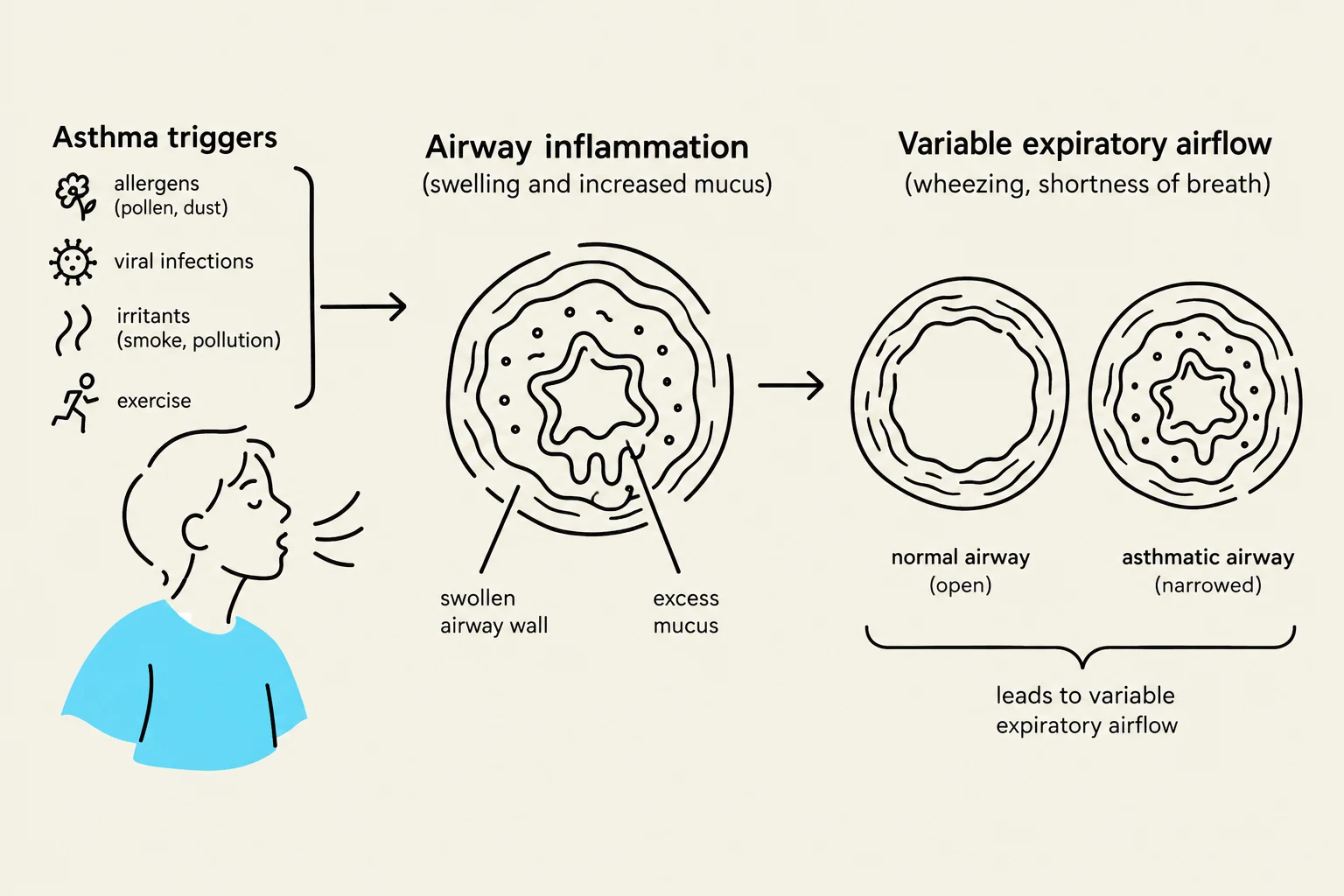
Asthma is a chronic condition characterized by two primary features: a history of varying respiratory symptoms and variable expiratory airflow limitation. Patients often report a constellation of symptoms, including wheezing, shortness of breath (dyspnea), chest tightness, and a cough that fluctuates in both timing and intensity.
A key diagnostic component is the use of a peak flow meter to measure the volume of air a person can exhale quickly after a full inspiration. In patients with asthma, these measurements correlate directly with airflow limitations.
Clinical Tip: When assessing a patient for an asthma exacerbation, listen for scattered expiratory wheezes during auscultation. Note that symptoms may be triggered by environmental factors such as cigarette smoke.
This diagram illustrates the process of asthma, starting with common triggers like allergens, viral infections, irritants, and exercise. It shows how these triggers lead to airway inflammation, characterized by swelling and increased mucus. Finally, it depicts the resulting variable expiratory airflow, comparing a normal, open airway to a narrowed asthmatic airway, which causes symptoms like wheezing and shortness of breath.
Bronchiolitis and Viral Infection
Bronchiolitis is an inflammation of the small airways (bronchioles) that is particularly significant in the pediatric population. The most frequent cause of this disorder is the respiratory syncytial virus (RSV).
Infants are at a higher risk for severe complications, especially those with a history of prematurity (specifically those born before 29 weeks of gestation). These preterm infants face an increased risk of both bronchiolitis and associated apnea. During a physical exam, clinicians should look for signs of respiratory distress, such as suprasternal, substernal, and intercostal retractions. These retractions occur because the infant must use accessory muscles to compensate for lung immaturity and airway obstruction.
Cystic Fibrosis: A Genetic Obstruction
Unlike asthma or bronchiolitis, Cystic Fibrosis (CF) is an inherited autosomal recessive disorder. It is caused by a mutation in the cystic fibrosis transmembrane conductance regulator (CFTR) protein.
The CFTR protein is responsible for the transport of chloride, sodium, and bicarbonate across cell membranes. When this protein malfunctions, it creates a physiological imbalance that leads to several systemic issues:
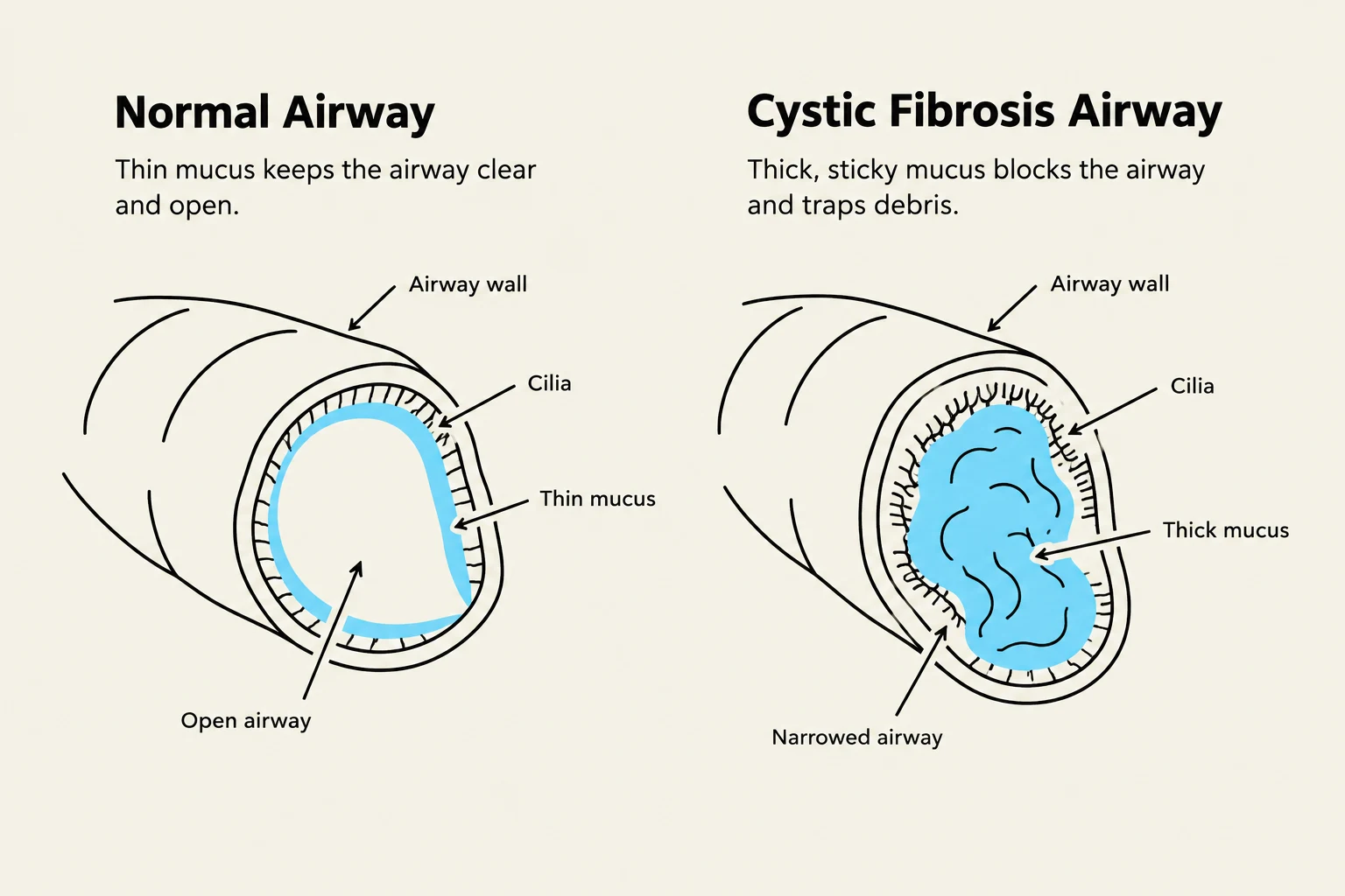
The production of thick, viscous mucus that obstructs the lungs, pancreas, liver, reproductive tract, and intestines.
Increased sodium content within the sweat glands.
Significant structural changes and scarring within the airways and lung tissue.
Comparison of a normal airway with thin mucus and cilia, versus a cystic fibrosis airway with thick mucus that narrows the passage.
Obstructive Sleep Apnea (OSA)
Obstructive Sleep Apnea (OSA) is a disorder of sleep-related breathing characterized by repeated episodes of reduced airflow or the temporary complete cessation of airflow during sleep.
The underlying pathology involves the physical collapse of the upper airway during sleep. This collapse necessitates an increased work of breathing, which triggers a brief arousal to activate the upper airway muscles, allowing the passage to reopen.
Pneumonia: Lower Respiratory Infection
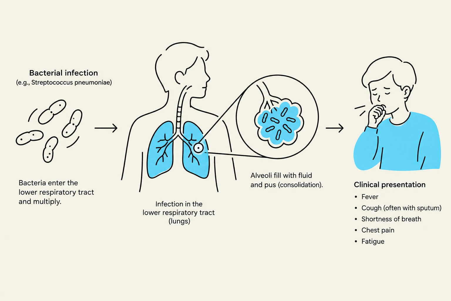
Pneumonia is a bacterial infection of the lower respiratory tract. It is clinically identified through a triad of findings: a cough, abnormal vital signs (such as fever or tachypnea), and abnormal lung examination findings.
Physical examination findings for pneumonia may include:
Dullness to percussion over the affected area.
Increased tactile fremitus (vibrations felt on the chest wall).
Auscultatory findings such as crackles (rales).
Imaging plays a vital role in confirming the diagnosis. For example, a chest X-ray may reveal signs of early pneumonia in a specific lobe, such as the left lower lobe.
Diagram illustrating the process of bacterial pneumonia, from bacterial entry into the respiratory tract to infection of the lungs and clinical symptoms.