This guide details the systematic clinical examination of the eye, focusing on observations of the iris, pupil, and conjunctiva. It covers normal characteristics, common abnormalities like conjunctivitis and subconjunctival hemorrhage, and their potential implications for systemic health.
Clinical examination of the eyes requires a systematic approach, moving from external structures to the internal fundus. By observing the iris, pupil, conjunctiva, and retina, clinicians can identify localized ocular pathologies as well as systemic conditions such as hypertension, diabetes, or Graves' disease.
External Examination: Iris and Pupil
The first step in a detailed ocular exam involves inspecting the iris and the pupil. The iris pattern should be clearly visible to the clinician. Under normal physiological conditions, both irides are expected to be the same color.
A critical component of this assessment is the evaluation of the pupils. Normal pupils are characterized by being round, regular, and equal in size. Clinicians must note any deviations from these characteristics, whether they occur unilaterally (in one eye) or bilaterally (in both eyes). For example, enlarged pupils may be a clinical sign of congenital glaucoma, while a "keyhole pupil" represents a specific structural abnormality.
Clinical Tip: Assessing the Anterior Chamber
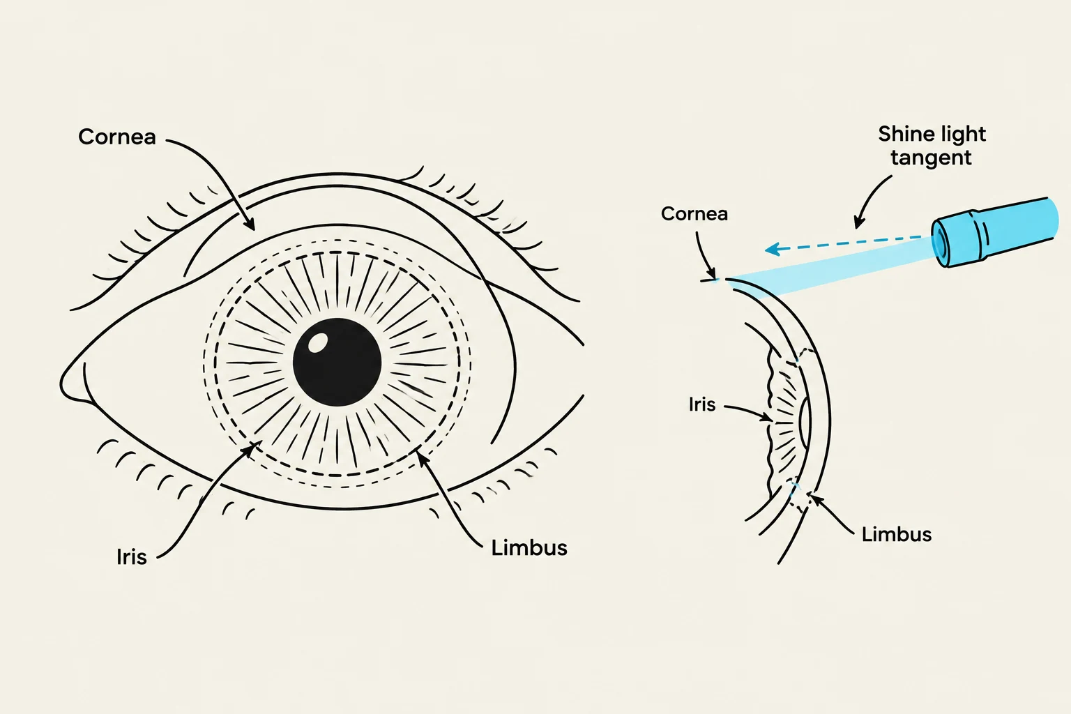
Before instilling mydriatic agents (which dilate the pupil), always inspect the patient's anterior chamber by shining a focused light tangentially at the limbus—the junction where the cornea meets the sclera. If the anterior chamber appears shallow, there is a significant risk of acute-angle glaucoma, and mydriatics should be avoided.
Diagram illustrating the cornea, iris, and limbus of the eye, with a depiction of shining a light tangent to the cornea.
Conjunctival Observations
The conjunctiva provides vital clues regarding infection, allergy, and vascular integrity. Clinicians should observe the color, texture, and presence of any discharge or blood.
Conjunctivitis and Inflammation
Inflammation of the tarsal conjunctiva is a common finding. Depending on the clinical presentation, this may indicate:
Allergic conjunctivitis: Often associated with itching and specific patterns of inflammation.
Infectious conjunctivitis: May present with significant redness (hyperemia) and the accumulation of thick, yellowish-white purulent exudate in the inner corner (canthus) and along the eyelid margins.
Subconjunctival Hemorrhage
A subconjunctival hemorrhage appears as a patch of bright red blood in a sharply defined area, surrounded by healthy-appearing conjunctiva. This blood remains bright red because of the direct diffusion of oxygen through the conjunctiva.
Triggers for these hemorrhages include:
Physical strain: Such as violent coughing, powerful sneezing, or straining during bowel movements.
Physiological states: Spontaneous occurrence during pregnancy or labor.
External factors: Trauma, foreign objects, or aggressive rubbing of the eye.
Most subconjunctival hemorrhages resolve spontaneously without intervention.
Ophthalmoscopy and Fundus Examination
To visualize the internal structures of the eye, clinicians perform ophthalmoscopy to examine the fundus.
The Red Reflex and Optic Disc
The examination begins by locating the red reflex, which is the light reflecting off the retina.
Opacities: Any obstructions in the path of light will appear as black densities within this reflex.
Absence of reflex: A missing red reflex may be due to improper equipment positioning, a cataract, or hemorrhage into the vitreous humor.
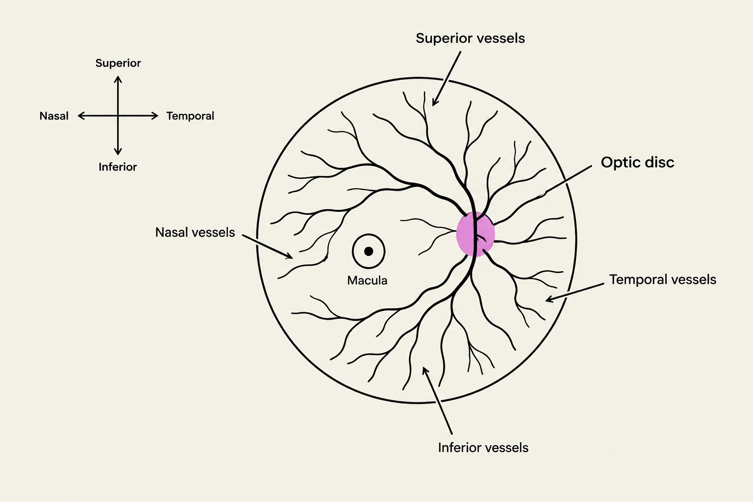
Once the red reflex is established, the clinician identifies the optic disc. While no discrete pigmentation is expected, small crescents may appear at the disc margin, most commonly along the temporal edge.
Diagram illustrating the anatomy of the retina, including the optic disc, macula, and major blood vessels.
Retinal Details and Vascularity
As the clinician approaches the retina, the blood vessels are typically the first structures to become visible. The vessels branch from the optic disc into superior, inferior, nasal, and temporal directions.
Clinicians should pay close attention to the characteristics of the vessels, as they can indicate systemic health:
Hypertension: May present as narrowing of the vessels, increased vascular tortuosity, "copper wiring" (a diffuse red-brown reflex), or arterovenous nicking.
Diabetes: Often associated with changes in corneal sensitivity (via the trigeminal nerve) and potential retinal complications.
Aging and Macular Degeneration
As individuals age, certain physiological changes occur. In older adults, lacrimal gland involution leads to decreased tear production. The most frequent causes of decreased visual function in this population are glaucoma, cataracts, and macular degeneration.
Drusen and the Amslar Grid
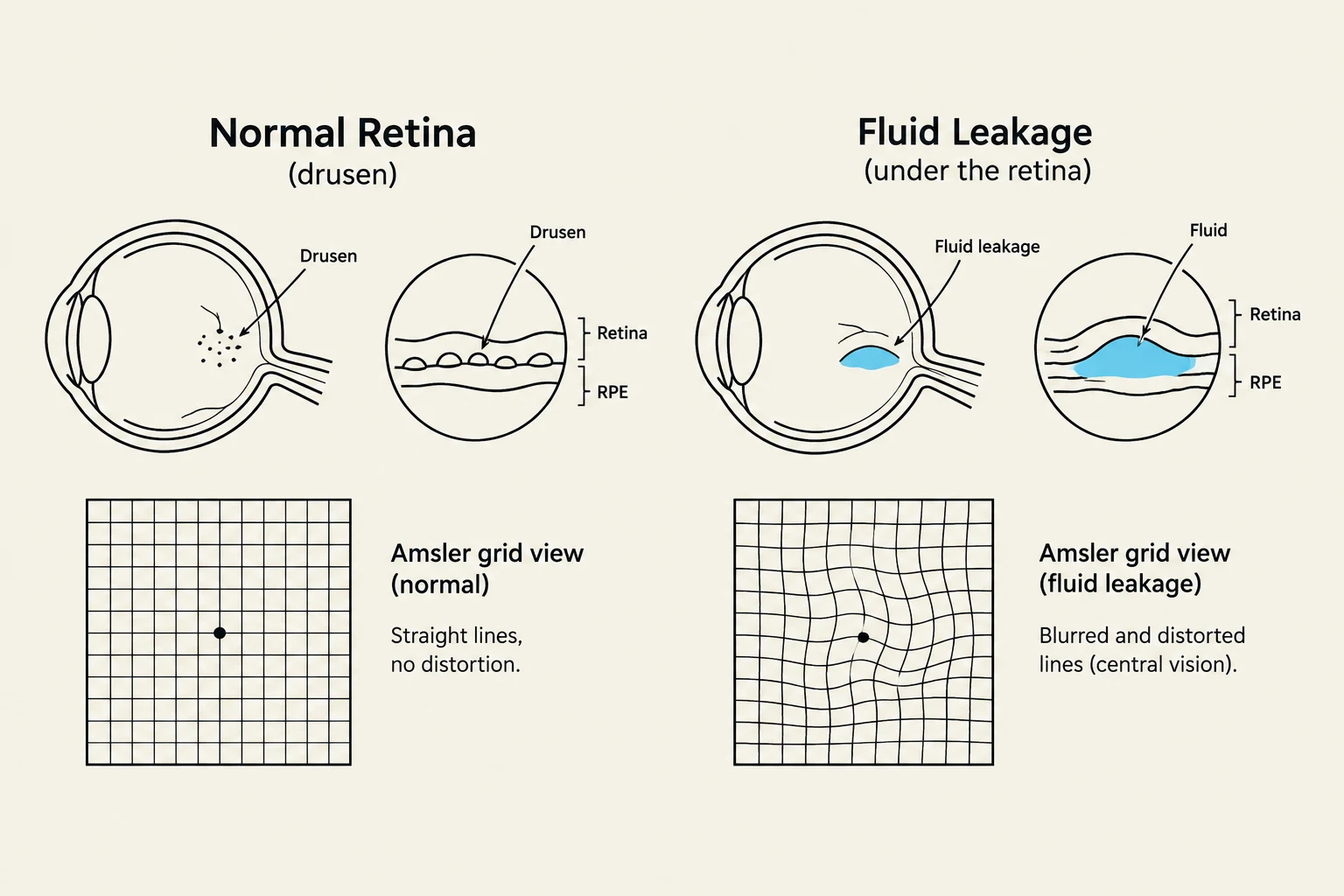
Drusen bodies are small, discrete, yellowish-white spots on the retina. While they can be a normal part of the aging process, an increase in their number or the intensity of their color can serve as a precursor to senile macular degeneration.
When drusen are noted, clinicians use the Amslar grid to evaluate the patient's central vision. The patient observes the grid to check for distortion (wavy or broken lines), which indicates macular changes or fluid leakage.
Diagrams illustrating a normal retina with drusen and fluid leakage under the retina, alongside corresponding Amsler grid views showing normal vision and distorted vision due to fluid leakage.
Clinical Precautions for Retinal Edema
In cases where macular degeneration leads to fluid leakage under the retina, the tissue may take on a glistening appearance indicative of edema.
Clinical Warning: Systemic Absorption
If retinal edema is present, clinicians should avoid the use of cycloplegic and mydriatic agents to prevent systemic absorption, unless they are absolutely necessary for the evaluation. To mitigate the risk of systemic absorption of topical medications, nasolacrimal duct occlusion can be employed.
Keep reading for free.
Create an account to finish this reading and make it part of your study routine.