


When a patient presents with an oral lesion, the clinician's task is to reconstruct a timeline and build a diagnostic framework that moves from observation to definitive management. Evaluating an intraoral lesion is not merely about identifying what is visible; it is about understanding the sequence of events, the systemic context of the patient, and the potential for serious underlying pathology.
Understanding Lesion Morphology
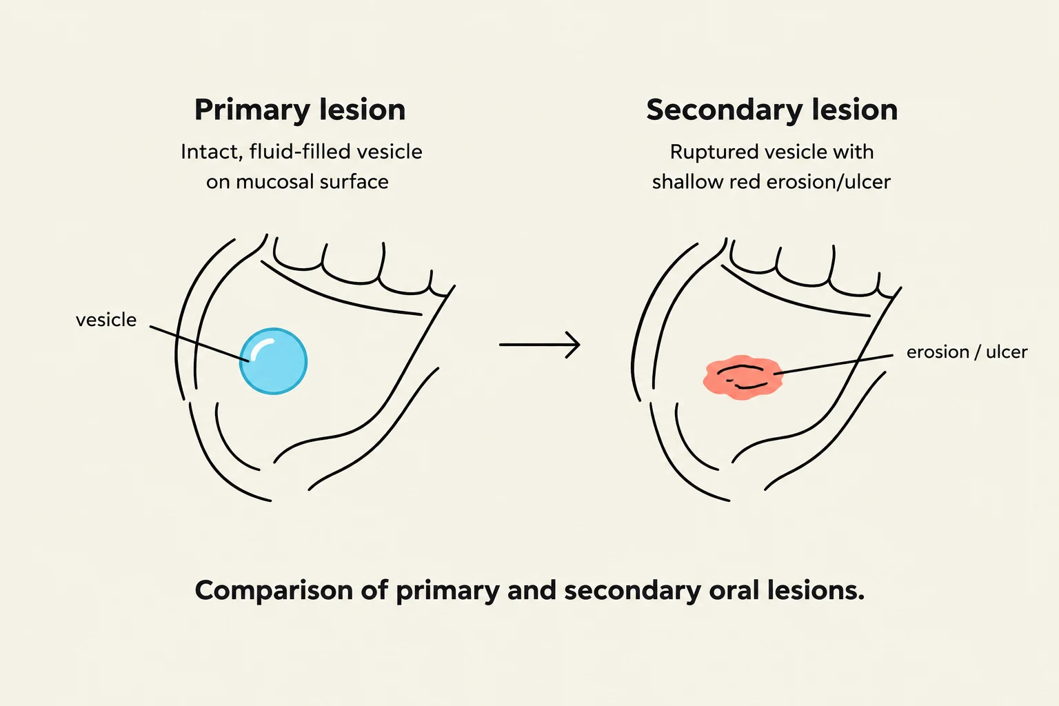
A secondary lesion occurs when the primary lesion is altered by external forces or natural progression. For example, if a vesicle ruptures, the resulting surface erosion or ulcer is considered a secondary lesion. It is crucial to recognize that trauma can significantly alter the original appearance of a lesion, potentially masking the primary cause.
The Diagnostic Process: History and Examination
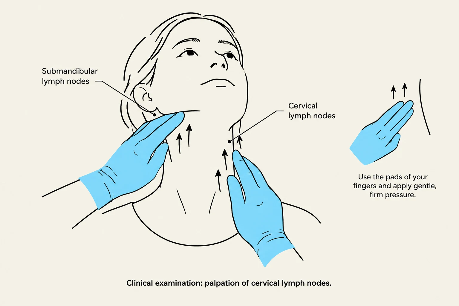
Focused Lesion History
To reconstruct the sequence of the lesion, you must perform a "focused lesion history". Rather than asking general questions, you should dive into the specifics of the lesion itself:
- Onset and Evolution: When did it begin? How has it changed over time?
- Symptoms: Is there pain, bleeding, or functional impairment (e.g., difficulty eating or needing to avoid hot/spicy foods)?
- Pattern of Occurrence: Does the lesion recur in the same spot, or does it appear in different locations? This helps differentiate between conditions like recurrent herpes and other chronic issues.
- Exposures: Review local trauma, medications, and recent dietary changes (e.g., new toothpaste, cinnamon-flavored foods, or even extremely hot food like pizza cheese).
Keep reading for free.
Create an account to finish this reading and make it part of your study routine.
Continue reading freeAlready have an account? Log in
Updated