This guide details the clinical assessment of the integumentary system, including skin, hair, and nails. It covers subjective data collection, inspection and palpation techniques for color, temperature, texture, and turgor, identification of primary lesions and melanoma using the ABCDE criteria, and assessment of hair
The integumentary system—comprising the skin, hair, and nails—serves as the body's first line of defense, providing protection, temperature regulation, and sensory perception. A clinical assessment of these structures is more than a superficial check; it is a window into a patient's systemic health, capable of revealing underlying issues such as anemia, liver disease, or chronic hypoxia. By combining systematic inspection and palpation with an understanding of biocultural variations, clinicians can provide accurate diagnoses and effective health promotion.
Preparing for Assessment and Subjective Data
Before beginning a physical exam, the clinician must control external variables such as room temperature and lighting—natural daylight is ideal. It is also vital to understand the patient's baseline; if it is a first visit, always ask about their usual skin color.
Assessment begins with subjective data collection. This involves interviewing the patient about their health history to identify potential risks or chronic conditions. Key questions should focus on:
Changes in pigmentation or moles: To screen for malignancy.
Skin sensations: Such as pruritus (itching) or excessive dryness (xerosis).
Environmental/Occupational hazards: Including sun exposure and chemical contact.
Hair and nail changes: To investigate potential nutritional deficiencies or endocrine issues.
Clinical Tip: When assessing patients with darker skin tones, do not rely solely on surface color. Inspect areas with less pigment, such as the sclera, oral mucosa, and nail beds, to identify systemic changes like jaundice or cyanosis.
Skin Inspection: Color and Pigmentation
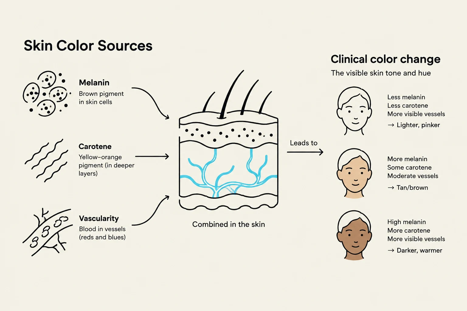
The primary goal of inspection is to evaluate color, pigmentation, and the presence of lesions. Skin color is determined by three sources: melanin (brown), carotene (yellow-orange), and the underlying vascular bed (red-purple).
Clinicians must be aware of biocultural variations to avoid misdiagnosis. For example, "alcohol flush syndrome" causes facial redness in some Indigenous and Asian populations and should not be mistaken for intoxication. Additionally, Inuit populations may exhibit different sweating patterns as an adaptation to extreme cold.
Identifying Color Changes
Widespread color changes can indicate serious physiological distress:
Pallor: A pale or ashen appearance often linked to anemia, shock, or arterial insufficiency.
Erythema: Redness caused by inflammation, fever, or cellulitis.
Cyanosis: A bluish tint indicating hypoxemia or poor perfusion.
Jaundice: A yellowing of the skin and sclera typically associated with liver disease.
Carotenemia: A yellow-orange tint to the palms and soles caused by excess carotene intake; unlike jaundice, the sclera remains white.
This diagram illustrates the sources of skin color, including melanin, carotene, and vascularity, and how their combination leads to different skin tones.
Palpation Techniques
Once inspection is complete, palpation is used to assess the physical properties of the skin.
Temperature, Moisture, and Texture
Temperature: Use the dorsa (backs) of your hands to compare both sides of the body. Abnormalities include localized hyperthermia (inflammation) or generalized hypothermia (shock).
Moisture: Note if the skin is excessively dry (xerosis) or if the patient is experiencing diaphoresis (profuse sweating).
Texture and Thickness: Skin should be smooth and even. Very thin, shiny skin may indicate aging or arterial insufficiency.
Mobility, Turgor, and Edema
To assess hydration, pinch a fold of skin under the clavicle or on the forearm.
Mobility refers to how easily the skin lifts.
Turgor is the skin's ability to return to its original position. "Tenting" (poor turgor) is a hallmark of dehydration.
Edema (fluid accumulation in interstitial tissue) is assessed by pressing firmly over a bony surface, such as the tibia, for five seconds. It is graded on a scale:
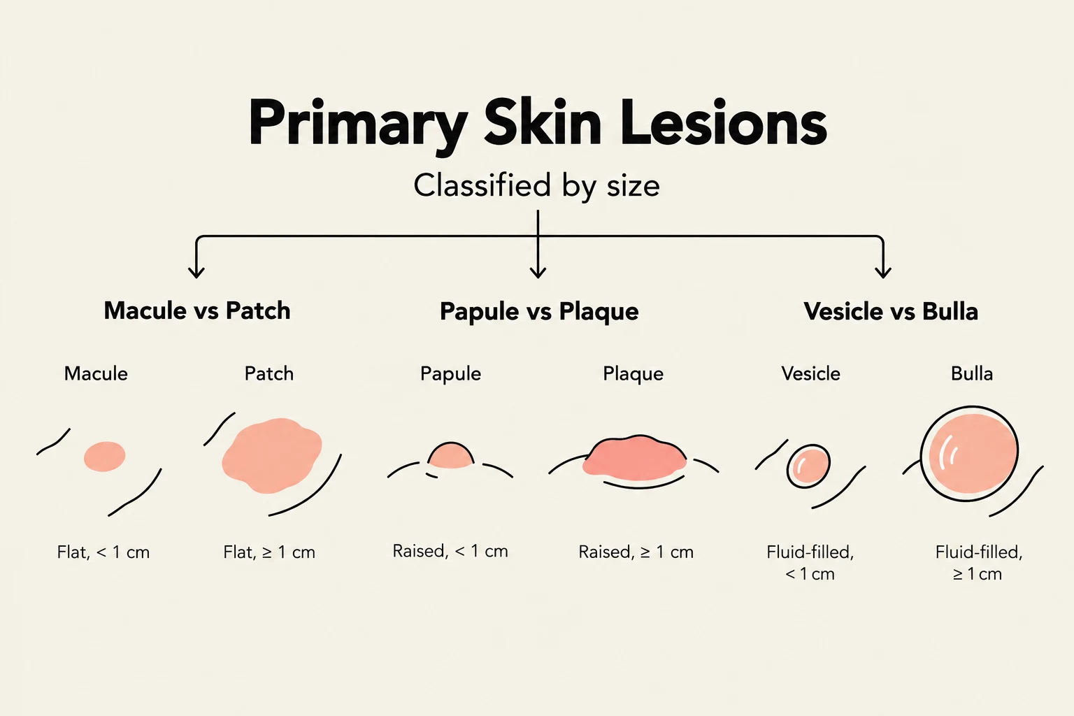
This diagram illustrates and classifies primary skin lesions based on their size and characteristics, differentiating between macules, patches, papules, plaques, vesicles, and bullae.
The ABCDEs of Melanoma
Because skin cancer is the most common cancer diagnosed in Canada, clinicians must use the ABCDE criteria to identify suspicious moles:
A - Asymmetry: One half does not match the other.
B - Border: Irregular, notched, or blurred edges.
C - Colour: Variegated (multiple colors or shades).
D - Diameter: Greater than 6 mm.
E - Evolution: Changing in size, shape, or symptoms (itching/bleeding).
Hair and Nail Assessment
Hair and Scalp
Inspection should include the scalp for lesions, flaking, or nits (lice eggs).
Alopecia: Patchy or diffuse hair loss.
Hirsutism: Excessive body or facial hair, often linked to endocrine disorders.
Texture: Brittle, dry hair may indicate hypothyroidism.
Nails
Nails are hard plates of keratin that appear pink due to the vascular nail bed.
Clubbing: An abnormal angle of ≥180∘ at the nail base, often indicating chronic hypoxia (e.g., lung or heart disease).
Capillary Refill: Press the nail edge to blanch it; color should return within 1–2 seconds. Delayed refill indicates poor peripheral perfusion.
Comparison of a normal nail profile with a 160° angle and a clubbed nail with a 180° angle, indicating chronic hypoxemia.
Health Promotion and Prevention
Preventative care is essential for reducing the incidence of skin disease and infections.
Sun Safety:
To minimize the risk of skin cancer, patients should follow the Canadian Cancer Society guidelines:
Check the UV index; protect skin when UV >3.
Seek shade between 11 a.m. and 3 p.m..
Use sunscreen with SPF 30+ and reapply every 2 hours.
Wear protective clothing and sunglasses with UVA/UVB protection.
Lyme Disease Prevention:
Lyme disease is a significant vector-borne threat in Canada. Prevention includes:
Wearing long sleeves and pants tucked into socks.
Using DEET repellent.
Performing daily full-body tick checks and removing ticks promptly with tweezers.
Note on Pressure Injuries: For bed-bound or chair-bound patients, prevention is far easier than treatment. Use the Braden Scale to assess risk; a lower score indicates a higher risk of developing pressure injuries.
Keep reading for free.
Create an account to finish this reading and make it part of your study routine.