This guide details the process of clinical observation, precise description of oral lesions, and targeted investigations for accurate diagnosis in oral medicine. It covers biopsy principles, avoiding false negatives, and the importance of follow-up and timely referrals to exclude malignancy.
In the field of oral medicine, the transition from observation to diagnosis is a critical bridge that requires meticulous precision. A clinician's ability to accurately describe a lesion and reason through a differential diagnosis can mean the difference between timely intervention and a missed diagnosis of malignancy. Effective management relies on the tight correlation between visual clinical findings and pathological results, ensuring that no "red flags" are overlooked during the diagnostic process.
Clinical Observation and Precise Description
A successful assessment begins with observation and interpretation. To support clinical reasoning, practitioners must move beyond vague terms and use approved morphological language to describe every lesion.
The Anatomy of a Description
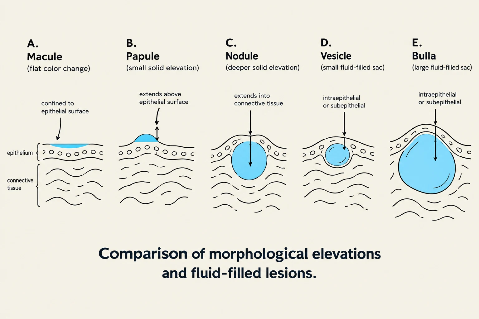
Comparison of morphological elevations and fluid-filled lesions, illustrating macules, papules, nodules, vesicles, and bullae.
When documenting a lesion, a clinician should record:
Site and Dimensions: Specify the exact anatomical sub-site (e.g., left buccal mucosa) and measure the longest dimension and perpendicular width in millimeters.
Number and Distribution: Is the lesion solitary, multiple, bilateral, or diffuse (scattered like sand)?
Color and Surface: Describe the color (white, red, pigmented, or variegated/speckled) and the surface texture (smooth, rough, reticular/lacy, or crusted).
Borders and Base: Note if the margins are well-defined or poorly defined, and if the base is pedunculated (on a stalk) or sessile.
Consistency and Fixation: Palpate to determine if the tissue is soft, fluctuant, firm, or indurated (hardened).
Morphology Vocabulary
This diagram compares superficial erosion, which affects only the epithelial layer, with deep ulceration, which involves a greater depth of tissue loss.
Using specific terms helps standardize communication between clinicians and pathologists:
Macule/Patch: A flat color change with no change in tissue elevation. A patch is simply a larger macule.
Papule/Nodule/Plaque: A papule is a small solid elevation; a nodule extends deeper into the connective tissue; a plaque is a broad, relatively flat elevation.
Vesicle/Bulla: These are fluid-filled sacs; a bulla is a larger version of a vesicle.
Exam Tip: Erosions vs. Ulcerations
To differentiate these, look at the depth of tissue loss. An erosion is a superficial loss of the epithelial layer that typically heals without scarring. An ulceration involves the crossing of the epithelium, reaching the connective tissue, and may leave a scar.
Targeted Investigations and Clinical Reasoning
Every investigation performed must be designed to answer a specific clinical question. Clinical reasoning involves using physical maneuvers to narrow the differential diagnosis.
Diagnostic Maneuvers
Scraping: Performed on white lesions to investigate potential Candida colonization. However, clinicians must remember that Candida can inhabit healthy tissue, so a positive scrape does not always indicate diseased tissue.
Stretching the Mucosa: This is a vital tool for differentiation. For example, stretching the cheek can help identify leukoedema if the white appearance diminishes or disappears. Conversely, stretching will not affect the appearance of linea alba.
The Importance of History
A patient's history reconstructs the sequence of events. Clinicians should ask about the onset, duration, pain, and functional effects (e.g., difficulty eating). Crucially, one must ask about systemic "red flags" such as unexplained weight loss, fatigue, or involvement of the eyes and genitals, which may point toward systemic syndromes.
Biopsy Principles and Malignancy Exclusion
When clinical suspicion is high, a biopsy is necessary to reach a definitive diagnosis. However, a biopsy is only as good as the sample provided.
Avoiding False Negatives
Diagram illustrating the correct and incorrect sites for an incisional biopsy, highlighting the risk of a false negative result if the biopsy is taken from an inappropriate area.
A common clinical reasoning error is assuming a negative biopsy result excludes malignancy. There are several reasons why this might occur:
Incorrect Sampling Site: In an incisional biopsy, the clinician must sample the most suspicious area. If the sample is taken from a clinically normal-looking area adjacent to dysplastic or cancerous tissue, the result will be a false negative.
Inadequate Sample Size: For large or complex lesions, taking multiple samples from different locations is recommended to ensure a representative view of the tissue.
Clinical Pearl: When to Re-evaluate
If a patient presents with systemic red flags (e.g., weight loss) or if the clinical appearance is highly suspicious for squamous cell carcinoma, do not accept a single negative biopsy as the final word. You should request the pathologist to revisit the slide or perform a new biopsy from a different site.
Urgency, Follow-up, and the Diagnostic Process
The diagnostic process does not end when the biopsy is taken; it ends when the patient is managed appropriately.
Timeliness and Referral
Certain conditions, such as untreated squamous cell carcinoma, can progress rapidly. Prompt specialist referrals (to oral surgeons or oral medicine specialists) are essential. When referring, ensure the recipient has the appropriate expertise for the specific task, such as performing a complex biopsy.
The "Two-Week Rule"
If a clinician removes a local traumatic factor—such as smoothing a sharp tooth—the patient must be brought back for a re-evaluation, typically within two weeks. This allows the clinician to see if the lesion has healed or if it persists, which would necessitate further investigation.
Closing the Loop
Follow-up is an indispensable part of the diagnostic process. Clinicians must ensure that:
Referrals are completed.
Results are communicated to the patient.
A management plan is enacted based on those results.
Keep reading for free.
Create an account to finish this reading and make it part of your study routine.