This document explains the classification of respiratory disorders into ventilation disorders, pleural conditions, and gas exchange problems, detailing their physiological defects, typical ABG findings, and common examples.
Understanding respiratory failure and oxygenation requires more than just knowing that a patient is "short of breath." To provide effective clinical intervention, one must be able to pinpoint exactly where the physiological breakdown is occurring. Respiratory dysfunction is not a monolithic entity; rather, it is a complex failure of specific mechanical or chemical processes. By categorizing these issues into ventilation disorders, pleural conditions, and gas exchange problems, clinicians can move from observing symptoms to diagnosing the underlying mechanism.
The Mechanics of Ventilation Disorders
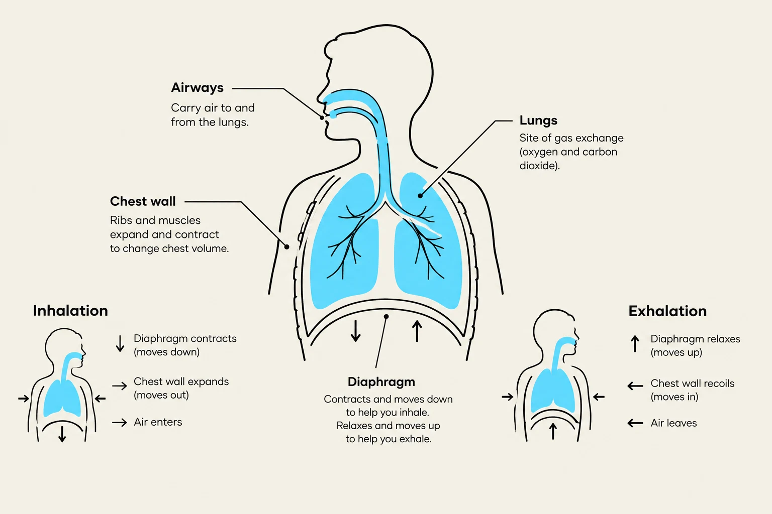
Ventilation is the mechanical process of moving air in and out of the lungs. It is a function of the "pump"—the muscles, the chest wall, and the airways. When we speak of a ventilation disorder, we are describing a failure of this pump to maintain adequate air movement, which leads to an accumulation of carbon dioxide (CO2) in the blood.
Ventilation disorders are often classified by their impact on the airway or the chest wall. For example, an airway obstruction (like asthma or COPD) increases resistance, making it harder to move air through the bronchial tubes. Conversely, a neuromuscular disorder (like Guillain-Barré syndrome or Myasthenia Gravis) represents a failure of the "engine" driving the pump. In both cases, the primary hallmark is hypercapnia—an elevation of partial pressure of carbon dioxide (PaCO2) in the arterial blood.
The relationship between ventilation and CO2 can be expressed by the simplified relationship:
Ventilation(V˙E)∝Metabolic RatePaCO2
If ventilation decreases while metabolic production of CO2 remains constant, PaCO2 must rise.
Diagram illustrating the process of human inhalation and exhalation, showing the roles of the diaphragm, chest wall, airways, and lungs.
Exam Tip: If a clinical vignette mentions high PaCO2 levels (hypercapnia), your first thought should be a ventilation problem. If the PaCO2 is normal or low, the problem is likely not with the mechanical movement of air, but with something else.
Pleural Conditions and Space-Occupying Issues
The lungs are housed within the pleural cavity, a potential space between the visceral pleura (covering the lungs) and the parietal pleura (lining the chest wall). For the lungs to expand fully, this space must maintain a negative pressure. When this space is compromised by the introduction of air, fluid, or blood, we encounter pleural conditions.
Pleural disorders can be broadly categorized into two types:
Pneumothorax: The presence of air in the pleural space, which causes the lung to collapse due to the loss of negative pressure.
Pleural Effusion: The accumulation of fluid (serous, blood, or pus) in the pleural space, which physically compresses the lung tissue and prevents full expansion.
While these conditions can eventually lead to gas exchange issues, their primary pathology is restrictive. They limit the volume of air the lungs can physically hold, regardless of how well the airways are functioning. A patient with a large pleural effusion may have perfectly clear airways, but they cannot take a deep breath because the lung is being squeezed by the surrounding fluid.
Diagrams illustrating normal lung function, pneumothorax (air in the pleural space), and pleural effusion (fluid in the pleural space).
Gas Exchange Problems: The Alveolar-Capillary Interface
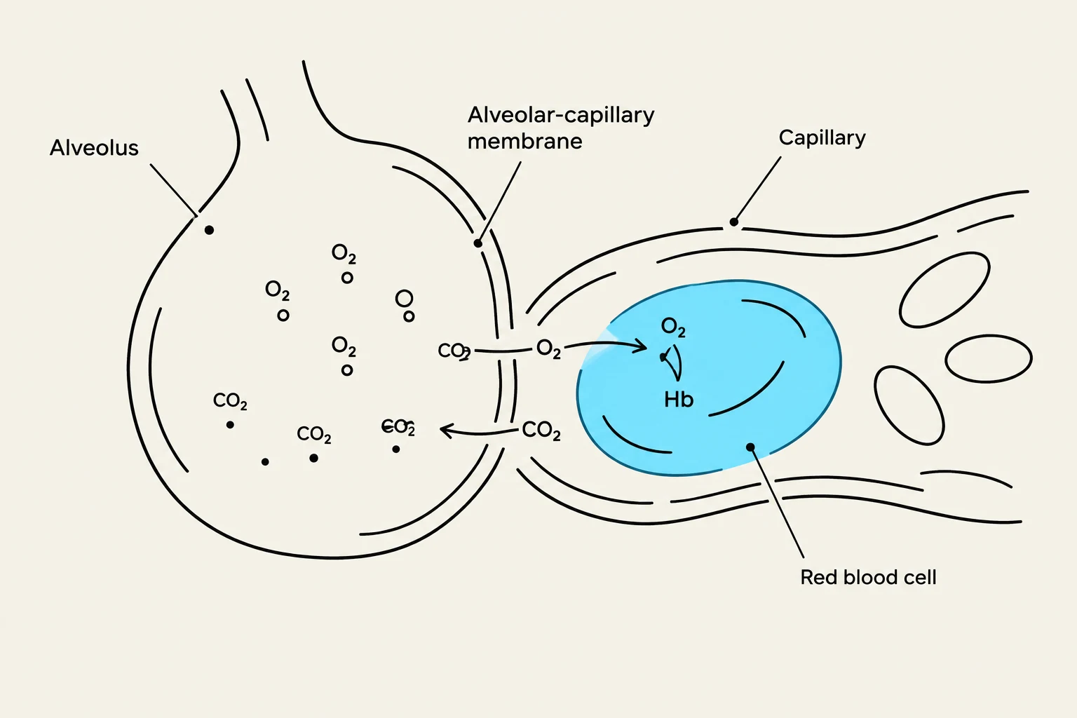
Even if ventilation is adequate (air is moving in and out) and the pleural space is clear (the lungs can expand), oxygenation can still fail. This occurs at the level of the alveolar-capillary membrane. This is the microscopic interface where oxygen moves from the air into the blood, and carbon dioxide moves from the blood into the air.
Gas exchange problems are typically driven by two main mechanisms:
V/Q Mismatch (Ventilation/Perfusion Mismatch): This occurs when the balance between the amount of air reaching the alveoli (V) and the amount of blood reaching the capillaries (Q) is disrupted. For example, in a pulmonary embolism, blood flow is blocked (low Q), meaning even if the lung is well-ventilated, no gas exchange can occur in that area.
Diffusion Impairment: This occurs when the membrane itself becomes thickened or damaged, making it physically difficult for gases to pass through. Conditions like pulmonary fibrosis increase the thickness of this membrane, slowing the rate of diffusion.
The efficiency of gas exchange is often represented by the PaO2/FiO2 ratio (the ratio of arterial oxygen partial pressure to the fraction of inspired oxygen). A low ratio is a hallmark of impaired gas exchange.
Diagram illustrating gas exchange between an alveolus and a capillary, showing oxygen (O2) entering the bloodstream and carbon dioxide (CO2) exiting.
Clinical Synthesis: Distinguishing the Disorders
To master this topic, you must learn to differentiate these categories using arterial blood gas (ABG) results and physical exam findings.
Disorder Category
Primary Physiological Defect
Typical ABG Finding
Common Examples
Ventilation
Failure of air movement (The Pump)
↑PaCO2 (Hypercapnia)
COPD, Opioid overdose, Myasthenia Gravis
Pleural
Restriction of lung expansion (The Space)
Variable (often ↓PaO2)
Pneumothorax, Pleural Effusion
Gas Exchange
Failure of oxygen/CO2 transfer (The Interface)
↓PaO2 (Hypoxemia)
Pulmonary Edema, Pulmonary Fibrosis, PE
The "Edge Case" Trap: Hypoxemia vs. Hypercapnia
Students often trip up by assuming that all respiratory problems cause high CO2. This is a mistake.
Consider a patient with a Pulmonary Embolism (PE). The lungs are being ventilated (air is moving), and the pleural space is clear. However, because the blood flow is blocked, gas exchange cannot occur. This patient will present with profound hypoxemia (low O2), but their CO2 might actually be low because they are hyperventilating in an attempt to compensate.
Conversely, a patient in respiratory arrest due to drug overdose has a ventilation problem. They aren't breathing enough, so CO2 builds up rapidly. The CO2 rise is the primary driver of their clinical decline.
By categorizing the problem into the Pump (Ventilation), the Space (Pleural), or the Interface (Gas Exchange), you can navigate even the most complex respiratory presentations.
Keep reading for free.
Create an account to finish this reading and make it part of your study routine.