


Cartilage is a specialized type of connective tissue that provides structural support, flexibility, and cushioning throughout the body. Unlike many other tissues, cartilage is unique because it lacks nerve endings, blood vessels (vasculature), and lymphatics. Because it is avascular, it relies on diffusion for nutrient delivery, which significantly impacts how thick cartilage can grow and how well it heals.
The Composition of Cartilage
As a connective tissue, cartilage is defined by its specific cellular components and its complex extracellular matrix (ECM). The ECM is not merely "filler"; it is a highly organized environment consisting of fibers and a gel-like ground substance that allows the tissue to resist compression.
The Cellular Components: Chondroblasts and Chondrocytes
The life cycle of a cartilage cell involves two main stages:
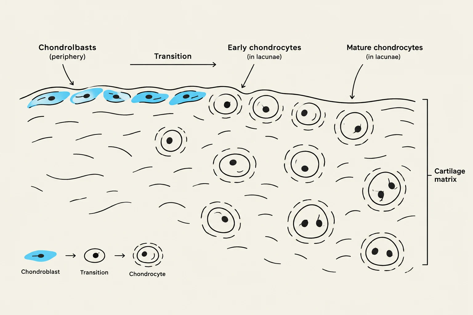
- Chondroblasts: These are found on the periphery of the cartilage in the chondrogenic zone. Their primary role is to secrete the matrix that forms the cartilage.
- Chondrocytes: As chondroblasts become embedded within the matrix they have created, they mature into chondrocytes. These mature cells reside in small spaces called lacunae. Once established, chondrocytes are responsible for maintaining the existing matrix.
The Extracellular Matrix (ECM)
The matrix is composed of two main elements: fibers and ground substance.
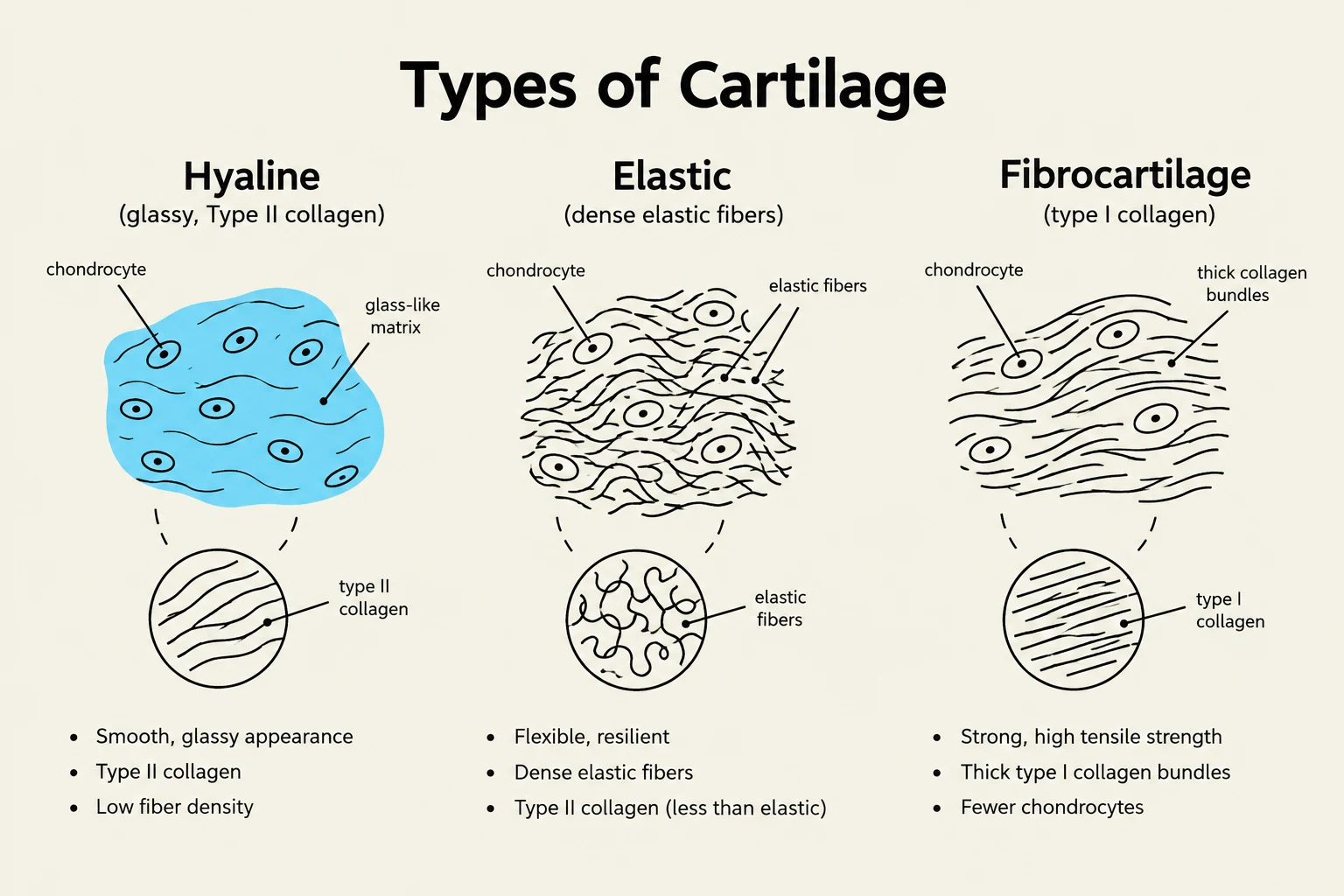
1. Fibers: Depending on the type of cartilage, the matrix contains collagen fibers (specifically Type II in hyaline and elastic cartilage) or elastic fibers.
2. Ground Substance: This is a highly hydrated, semi-solid substance that allows cartilage to act as a shock absorber. Its unique properties are driven by:
Keep reading for free.
Create an account to finish this reading and make it part of your study routine.
Continue reading freeAlready have an account? Log in
Updated