Explore chromosomal mutations, the process of meiosis, and the concept of aneuploidy. Understand how errors in cell division, such as nondisjunction, lead to changes in chromosome number and affect gene dosage, impacting organism development and health.
Chromosomal mutations and errors in cell division are not merely biological curiosities; they are fundamental drivers of evolution, critical factors in agricultural productivity, and significant contributors to human health and disease. While we often think of genetic variation in terms of single nucleotide changes, large-scale alterations to the structure or number of chromosomes can have profound effects on an organism's development and survival. This article explores the mechanisms behind these mutations, the intricacies of meiosis, and the concept of gene dosage that explains why having "too much" or "too little" of a chromosome can be so disruptive.
The Foundation: Meiosis and Genetic Variation
To understand chromosomal mutations, one must first understand the process of meiosis. Meiosis is the specialized cell division that transforms a diploid cell ($2n$) into haploid gametes (n). In humans, this means reducing the chromosome count from 46 to 23.
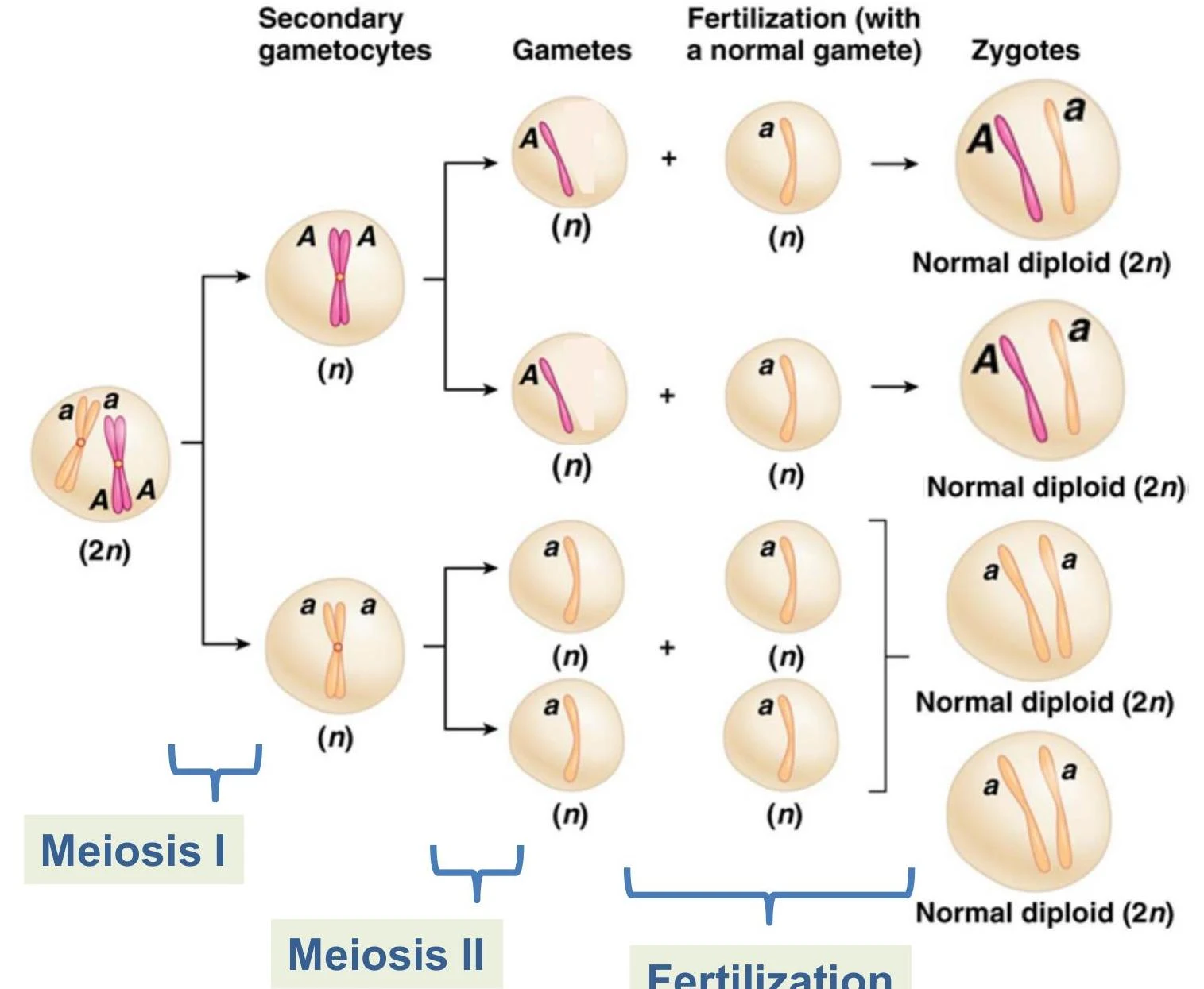
This diagram illustrates the process of meiosis and fertilization, showing how a diploid cell (2n) undergoes meiosis to produce haploid gametes (n), which then combine during fertilization to form diploid zygotes (2n).
Meiosis is not just about reduction; it is the engine of genetic diversity. Two primary mechanisms ensure that every gamete is unique:
Crossing Over (Recombination): During prophase I, homologous chromosomes exchange segments of DNA. This reshuffles the alleles on a single chromosome, creating new combinations that did not exist in the parents.
Independent Assortment: During metaphase I, homologous pairs align randomly at the cell equator. The direction in which a maternal or paternal chromosome faces is independent of other pairs, leading to a vast array of possible chromosomal combinations in the resulting gametes.
When a haploid gamete successfully fuses with another during fertilization, it forms a zygote, restoring the diploid number.
Chromosomal Mutations: Structural Changes
Chromosomal mutations can involve changes in the structure of the chromosomes themselves. These are often distinct from changes in the total number of chromosomes.
Duplications: A segment of a chromosome is repeated, increasing the copy number of specific genes.
Deletions: A segment of a chromosome is lost, which can lead to the loss of essential genetic information.
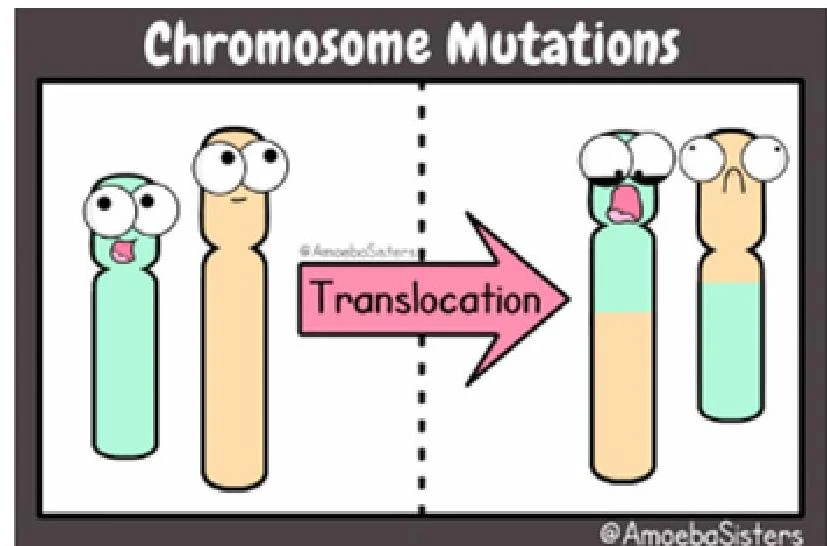
Translocations: This occurs when segments of two different, non-homologous chromosomes are swapped.
This diagram illustrates chromosome translocation, a type of mutation where a segment of one chromosome breaks off and attaches to another chromosome.
Aneuploidy and Nondisjunction
While structural mutations change the "map" of a chromosome, aneuploidy refers to a change in the number of individual chromosomes within a cell. This is distinct from polyploidy (or euploidy), where the entire genome is multiplied (e.g., triploidy, $3n$).
The Mechanism: Nondisjunction
Aneuploidy is primarily caused by nondisjunction. Nondisjunction is the failure of homologous chromosomes (during meiosis I) or sister chromatids (during meiosis II) to separate properly during cell division.
If nondisjunction occurs during meiosis, the resulting gametes will have an abnormal number of chromosomes. When these gametes participate in fertilization, the resulting zygote will exhibit aneuploidy:
Trisomy (2n+1): The zygote has three copies of a particular chromosome instead of two.
Monosomy (2n−1): The zygote is missing one copy of a particular chromosome.
Nullisomy: An entire homologous pair is missing.
Exam Tip: Pay close attention to when nondisjunction occurs. Nondisjunction in Meiosis I involves the failure of homologous chromosomes to separate, whereas in Meiosis II, it involves the failure of sister chromatids to separate.
Gene Dosage and the Central Dogma
Why does an extra chromosome cause such severe developmental issues? The answer lies in the Central Dogma of molecular biology: DNA is transcribed into RNA, which is then translated into proteins.
In a normal wild-type organism, the gene dosage—the relative amount of a gene product (protein) produced by a cell—is precisely balanced. Because gene expression is tied to the number of DNA copies available, an increase in the copy number of a gene leads to an increase in the amount of its corresponding protein.
Developmental processes are highly sensitive to the interaction of gene products. If a mutation (like a duplication or trisomy) increases the amount of one protein but leaves its interacting partners at normal levels, the delicate biochemical balance required for embryonic development is disrupted. This imbalance often results in abnormal development or lethality.
Analogy: Think of baking. If you change the relative proportions of ingredients (like doubling the flour but keeping the sugar the same), the final product will be fundamentally different and likely ruined.
Factors Influencing Phenotypic Impact
Not all aneuploidies are created equal. The severity of the phenotype (the observable traits) depends on several factors:
1. Taxonomy (Organism Type)
Plants generally tolerate aneuploidy much better than animals. This is due to different cellular mechanisms for managing dosage, more flexible body plans, and the ability to self-fertilize, which can help stabilize a population. For example, trisomy in Jimson weed is relatively common and mild, whereas monosomy in the evening primrose decreases fitness.
2. The Specific Chromosome
The "content" of the chromosome matters significantly:
Size and Gene Count: Aneuploidies involving smaller chromosomes (with fewer genes) are often better tolerated than those involving large chromosomes.
Sex Chromosomes: Humans tolerate aneuploidy in sex chromosomes (like Turner syndrome, XO, or Klinefelter syndrome, XXY) better than in autosomes because of existing mechanisms to balance gene dosage.
Autosomes: Trisomy of autosomes (non-sex chromosomes) is often severe. In humans, Trisomy 21 (Down syndrome), Trisomy 18 (Edwards syndrome), and Trisomy 13 (Patau syndrome) are well-known examples.
3. Cell Type: Germ Line vs. Somatic Cells
Germ Line Mutations: If nondisjunction occurs during meiosis, the error is present in every cell of the resulting offspring.
Somatic Mutations: If nondisjunction occurs during mitosis in a growing organism, only the descendants of that specific cell will be aneuploid. In humans, aneuploidy in somatic cells is a hallmark of many cancer cells.
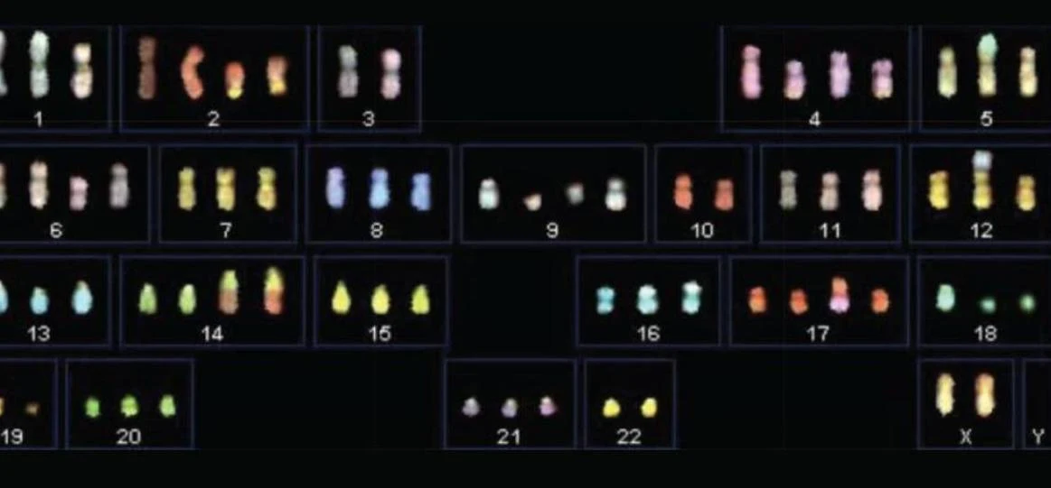
A karyotype showing homologous chromosomes arranged in pairs and numbered 1 through 22, along with the sex chromosomes X and Y.
Summary Table: Aneuploidy vs. Euploidy
Term
Type of Change
Description
Example
Aneuploidy
Numerical (Individual)
Change in the number of a single homologous pair
Trisomy 21, Monosomy X
Euploidy
Numerical (Whole Genome)
Change in the number of entire sets of chromosomes
Triploidy ($3n$), Tetraploidy ($4n$)
Keep reading for free.
Create an account to finish this reading and make it part of your study routine.