


Cervical cancer management is a highly specialized field that balances oncological safety with the preservation of quality of life and reproductive potential. Modern treatment protocols are increasingly driven by precise staging, advanced surgical techniques, and a deep understanding of tumor biology. This article serves as a comprehensive guide to the current standards in staging, surgical modalities, and clinical management for cervical cancer, primarily based on the NCCN Guidelines.
Staging and Diagnostic Protocols
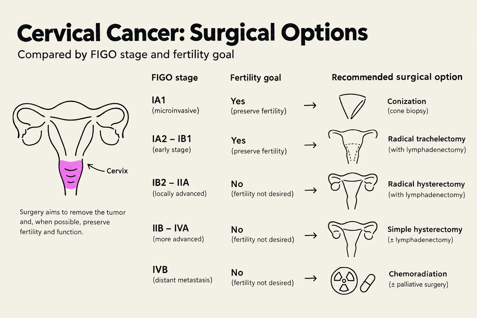
Accurate staging is the cornerstone of cervical cancer management. Current clinical practice is guided by the Revised FIGO staging system, which has evolved to incorporate more precise pathological and imaging findings. The management pathway for a patient is determined by their specific stage, ranging from microinvasive disease to locally advanced or metastatic presentations.
Pathological Assessment
The diagnostic process begins with a thorough pathological examination. Standards for examining specimens are defined by the College of American Pathologists (CAP) to ensure consistency in reporting. For specific histologies, such as endocervical adenocarcinoma, pathologists look for specific patterns, such as the "Silva pattern" of invasion, which is particularly relevant in HPV-associated cases.
Lymph Node Staging
One of the most significant shifts in recent years is the move toward minimizing surgical morbidity through improved lymph node assessment.
- Sentinel Lymph Node (SLN) Biopsy: For early-stage cervical cancer, SLN biopsy is a critical tool used to avoid the extensive morbidity associated with full pelvic lymphadenectomy. Techniques such as lymphoscintigraphy and the use of dyes like Indocyanine Green are employed to map the drainage.
- Para-aortic Lymphadenectomy: In more advanced or high-risk cases, clinicians must decide if para-aortic lymphadenectomy is necessary, often guided by the results of advanced imaging like PET.

Keep reading for free.
Create an account to finish this reading and make it part of your study routine.
Continue reading freeAlready have an account? Log in
Updated