Explores the brain's high metabolic demand, the critical importance of uninterrupted blood flow, and the consequences of disruptions like ischemic penumbra and cerebrovascular accidents (CVAs). Details the carotid and vertebral-basilar arterial systems, the Circle of Willis, and collateral circulation pathways.
The human brain is a metabolic powerhouse. Although it accounts for only 2% of total body weight, it consumes a staggering 20% of the body's total cardiac output. Because brain cells possess no internal energy storage mechanisms, they require a constant, uninterrupted supply of blood to deliver essential nutrients and remove metabolic waste products, such as carbon dioxide. This delicate balance makes the brain exceptionally vulnerable to any disruption in its vascular supply.
Blood Flow Disruption and the Ischemic Penumbra
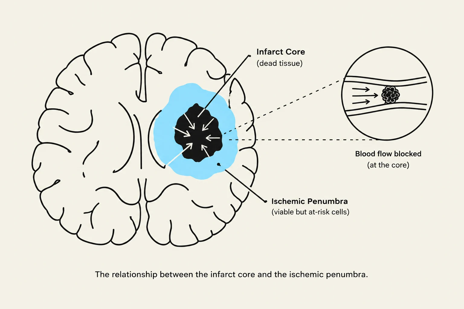
The relationship between the infarct core and the ischemic penumbra.
When blood flow is interrupted, the consequences are rapid and often devastating. Clinical thresholds for damage are very specific: a reduction in blood flow volume to below 20 mL/100g/min can result in irreversible damage to brain cells. Furthermore, a total vascular interruption lasting only 4–6 minutes can cause catastrophic, irreversible injury.
In the event of an occlusion, clinicians look for the ischemic penumbra. This refers to the area of "idle" but potentially viable cells surrounding the central site of an infarct (the dead tissue). The survival of these cells depends heavily on time and circulation:
Without alternate circulation: Cells in the penumbra may survive for approximately 20 minutes.
With adequate collateral supply: Cells may survive for 6–8 hours.
Exam Tip: When studying Cerebrovascular Accidents (CVAs), remember that 80% are related to arterial occlusion (ischemia), while only about 10% are hemorrhagic (bleeding).
Cerebrovascular Accidents (CVA)
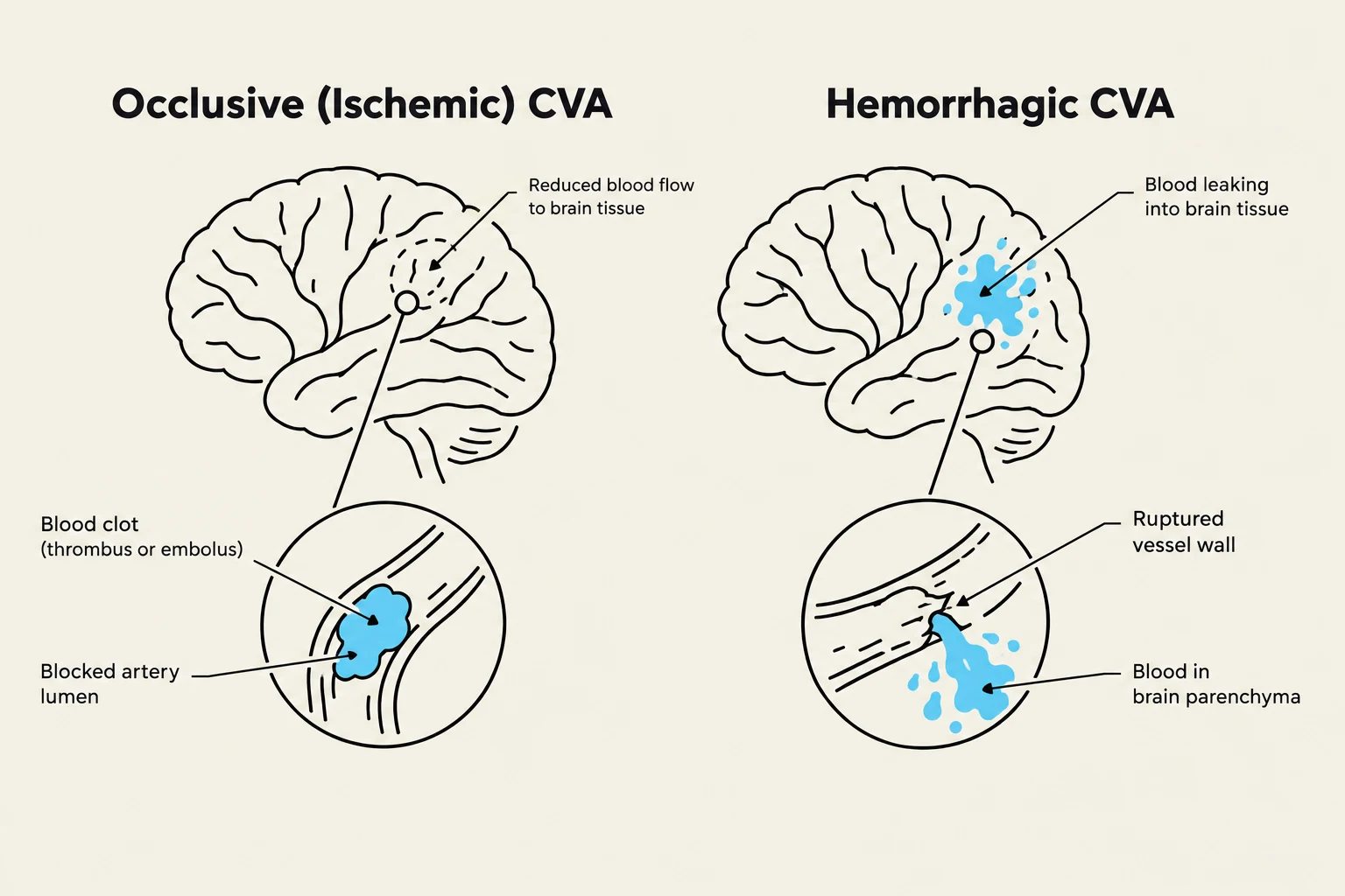
This illustration compares occlusive (ischemic) cerebrovascular accident (CVA) with hemorrhagic CVA. An ischemic CVA occurs when a blood clot blocks an artery, reducing blood flow to brain tissue. A hemorrhagic CVA occurs when a blood vessel ruptures, causing bleeding into brain tissue.
A CVA, or stroke, occurs when blood supply to a part of the brain is interrupted. These are generally categorized into three types:
Occlusive (Ischemic) CVAs: These account for the majority of cases and are further divided into:
Thrombosis (65%): A gradual build-up of plaque that occludes the artery over many years. These often occur during rest or sleep.
Embolism (25%): A blood clot that breaks free from a thrombus elsewhere and travels until it blocks a smaller end artery. These often affect younger people and occur during periods of activity.
Lacunar CVA: Involve small arteries of the basal ganglia and thalamus, often seen in patients with idiopathic hypertension.
Hemorrhagic CVAs (10%): Caused by the rupture of weakened blood vessel walls (such as an aneurysm or arteriovenous malformation) under the pressure of blood flow.
Transient Ischemic Attack (TIA): A temporary interruption of blood flow that does not result in permanent damage, though it serves as a significant warning sign; approximately one-third of people who experience a TIA later suffer a major stroke.
The Dual Arterial Systems
The brain receives its blood supply from two primary systems that eventually join at the base of the brain:
The Carotid System
Originating from the aorta, the carotid arteries ascend each side of the neck. They divide into two main branches:
External Carotid Artery: Supplies the facial muscles, forehead, and the oral, nasal, and orbital cavities.
Internal Carotid Artery: A major source of blood to the brain. It gives rise to the Anterior Cerebral Artery (ACA) and the Middle Cerebral Artery (MCA), and also includes branches like the ophthalmic artery, which supplies the eyes.
The Vertebral-Basilar System
Arising from the subclavian arteries, this system supplies the posterior and inferior structures of the brain. Before reaching the Circle of Willis, these arteries branch out to supply the spinal cord, medulla, pons, midbrain, and cerebellum. Key branches include:
Anterior Spinal Artery: Supplies the lower medulla. Occlusion can lead to "alternating hemiplegia" (ipsilateral facial/tongue paralysis and contralateral extremity paralysis).
Posterior Inferior Cerebellar Artery (PICA): Supplies a large portion of the cerebellum.
Anterior Inferior Cerebellar Artery (AICA): Includes the labyrinthine artery, which supplies the inner ear; involvement can cause deafness and vestibular dysfunction.
Superior Cerebellar Artery: Involvement can result in ataxia, impaired balance, and dysarthria.
The Circle of Willis and Collateral Circulation
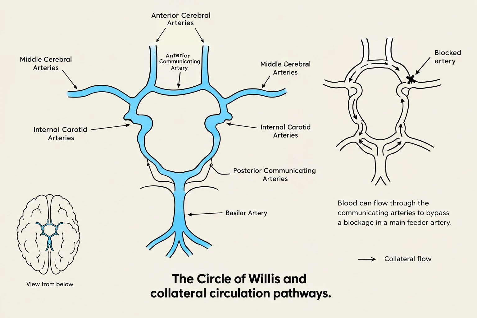
Diagram illustrating the Circle of Willis and collateral circulation pathways in the brain, showing how blood can reroute around a blockage.
At the ventral surface of the brain, the carotid and vertebral-basilar systems connect to form the Circle of Willis. This structure serves to equalize the vascular blood supply to both hemispheres. The circle contains both cortical arteries (supplying external structures) and central arteries (supplying internal/subcortical structures).
When a primary artery is blocked, the brain relies on collateral circulation—an alternative vascular supply. There are four primary points of potential collateral flow:
Anterior Communicating Artery: Links the two anterior cerebral arteries to connect the supply between both hemispheres.
Posterior Communicating Artery: Allows the vertebral-basilar system to feed the internal carotid system (or vice versa) if one becomes insufficient.
Ophthalmic Artery: Allows for retrograde (backward) flow from the external carotid to the internal carotid during thromboembolic events.
Watershed Areas: These are regions at the very end of an arterial distribution where the terminal branches of the three major cortical arteries meet or overlap. These areas are the most vulnerable to critically low cerebral blood flow.
Major Arterial Territories and Clinical Implications
The specific symptoms of a vascular event depend on which arterial territory is affected:
Artery
Primary Territories Supplied
Common Clinical Symptoms of Occlusion
Anterior Cerebral (ACA)
Medial cortical surfaces (prefrontal, frontal, parietal), corpus striatum, and internal capsule
Sensory loss and paralysis in the lower extremities; prefrontal lobe symptoms
Middle Cerebral (MCA)
Entire lateral surface of the brain, including sensorimotor and language areas
Contralateral hemiplegia, aphasia, tactile agnosia, and visual field changes
Posterior Cerebral (PCA)
Anterior/inferior temporal lobe and inferior/medial occipital lobe (including visual cortices)
Various visual field changes
Keep reading for free.
Create an account to finish this reading and make it part of your study routine.