Cellular Respiration: TCA Cycle and Electron Transport Chain
An explanation of aerobic cellular respiration, focusing on the TCA cycle (Krebs cycle) and the Electron Transport Chain (ETC) for ATP synthesis within mitochondria. Covers key enzymes, metabolic pathways, and the impact of toxins.
To understand how our bodies convert food into movement, thought, and warmth, we must look deep inside the mitochondria. The process of aerobic cellular respiration is a sophisticated multi-step relay where electrons are stripped from fuel molecules and used to power the synthesis of Adenosine Triphosphate (ATP). This energy production relies on two interconnected systems: the TCA Cycle (also known as the Krebs Cycle) and the Electron Transport Chain (ETC).
The TCA Cycle: Harvesting Electrons
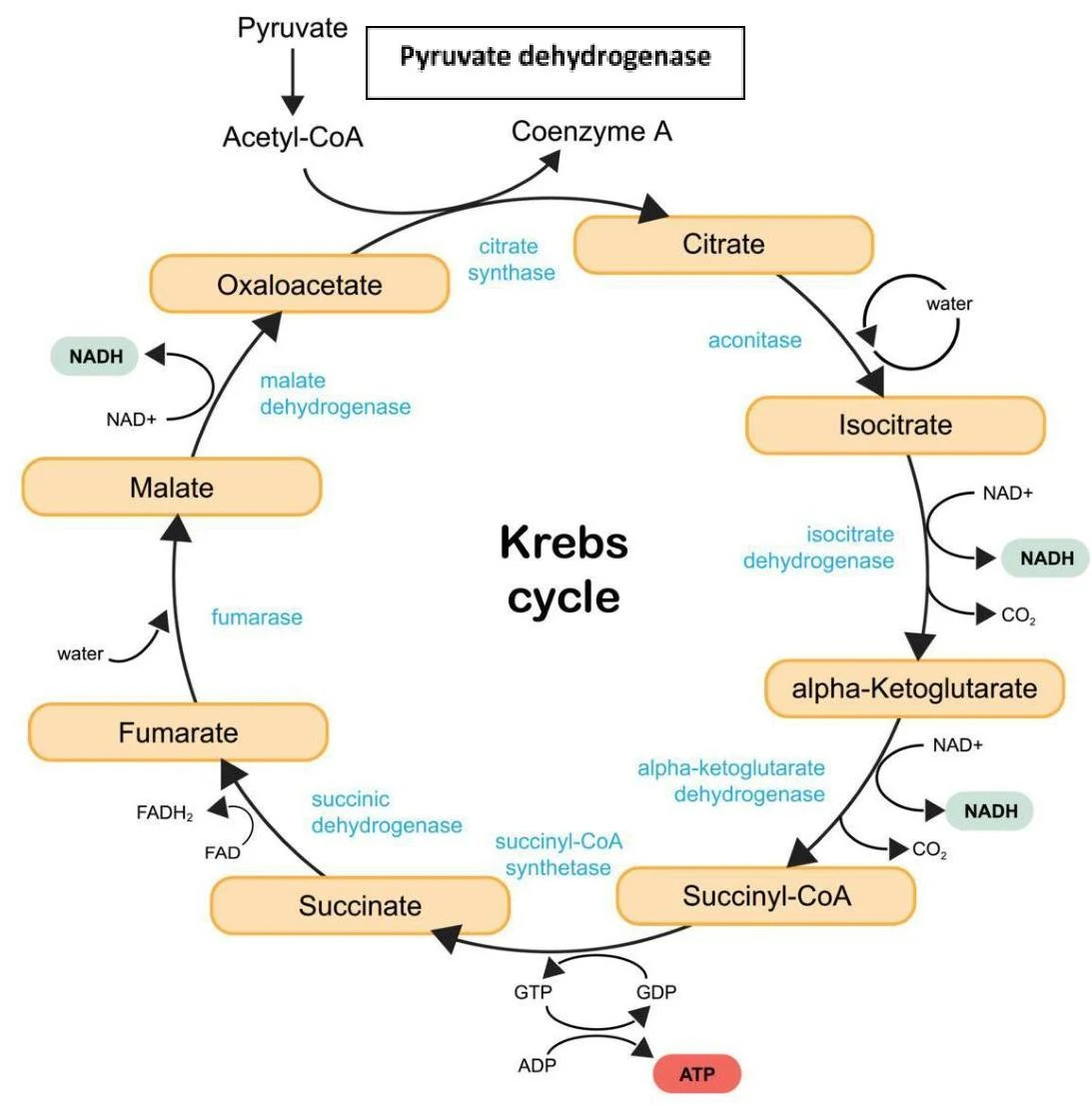
The TCA cycle is a series of enzymatic reactions occurring within the mitochondrial matrix. Its primary purpose is not to produce massive amounts of ATP directly, but to harvest high-energy electrons from acetyl-CoA and transfer them to carrier molecules.
For every one molecule of acetyl-CoA that undergoes complete oxidation, the cycle yields:
3 NADH
1 FADH2
1 GTP (which is energetically equivalent to ATP)
2 CO2 (released as a byproduct)
The Krebs cycle, also known as the citric acid cycle or TCA cycle, is a series of chemical reactions used to generate energy through the oxidation of acetyl-CoA derived from carbohydrates, fats, and proteins.
Key Regulatory Enzymes
The speed of the TCA cycle is tightly controlled by the cell's energy needs. If the cell has plenty of energy, the cycle slows down; if energy is low, it accelerates.
Isocitrate Dehydrogenase: This is the rate-limiting step of the cycle. It produces α-ketoglutarate and is inhibited by high levels of NADH and ATP.
α-Ketoglutarate Dehydrogenase: This enzyme also produces NADH and is subject to feedback inhibition by high NADH levels.
Pyruvate Dehydrogenase (PDH): While technically the bridge between glycolysis and the TCA cycle, PDH is critical. It converts pyruvate into acetyl-CoA.
Exam Tip: If PDH is blocked, pyruvate cannot enter the TCA cycle as acetyl-CoA. To regenerate NAD+ and allow glycolysis to continue, the cell will convert the excess pyruvate into lactate, leading to lactic acidosis.
Nutritional Dependencies
The efficiency of these enzymes depends on specific vitamins. A deficiency in Vitamin B1 (thiamine) impairs both PDH and α-ketoglutarate dehydrogenase. Furthermore, Vitamin B2 (riboflavin) is essential for the function of succinate dehydrogenase (which produces FADH2).
The Electron Transport Chain (ETC) and Oxidative Phosphorylation
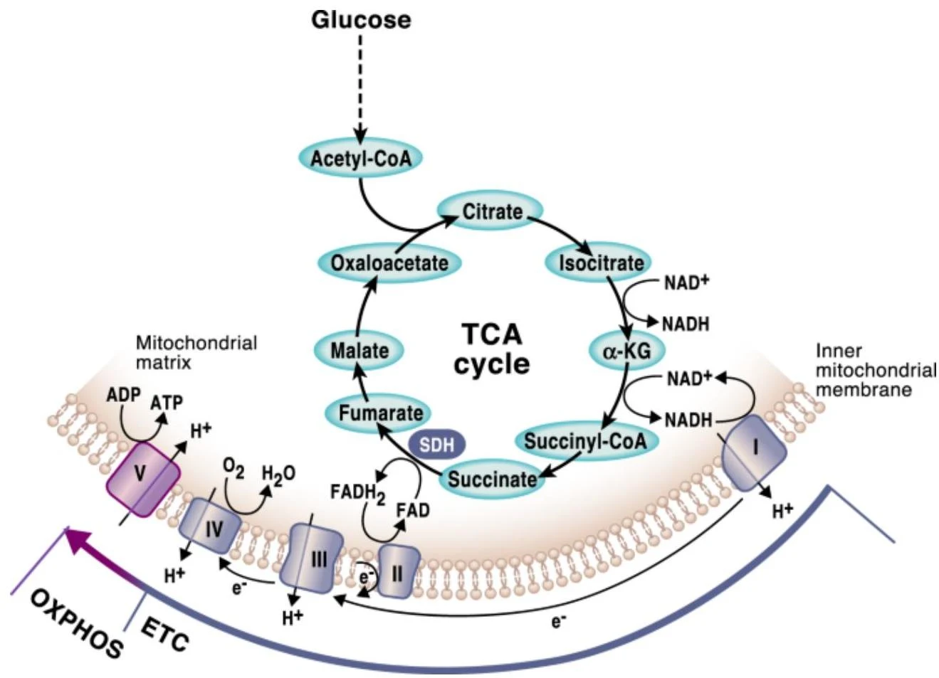
Once the TCA cycle has produced NADH and FADH2, these molecules travel to the inner mitochondrial membrane to participate in the Electron Transport Chain (ETC). The ETC is a series of protein complexes that use the energy from electrons to create a proton gradient.
Diagram illustrating the TCA cycle and oxidative phosphorylation within the inner mitochondrial membrane.
The Mitochondrial Complexes
The flow of electrons moves through several key components:
Complex I: Receives electrons from NADH.
Complex II (Succinate Dehydrogenase): Receives electrons from FADH2. Note that this enzyme is unique because it is a functional part of both the TCA cycle and the ETC.
Coenzyme Q (Ubiquinone): A critical mobile electron shuttle that carries electrons to Complex III.
Complex IV (Cytochrome c Oxidase): The final step where electrons are transferred to oxygen to form water.
Oxidative Phosphorylation and ATP Yield
The movement of electrons drives the pumping of protons ($H^+$) across the membrane, creating an electrochemical gradient. This gradient represents stored potential energy. The only way for these protons to flow back into the matrix is through Complex V (ATP Synthase). This process is called oxidative phosphorylation.
The energy yield from the electron carriers is approximately:
1 NADH ≈ 2.5 ATP
1 FADH2≈ 1.5 ATP
Consequently, one acetyl-CoA molecule yields a total of approximately 10 ATP equivalents (3 NADH × 2.5 + 1 FADH2× 1.5 + 1 GTP).
Metabolic Dysfunction and Toxicity
Because the ETC is the primary source of ATP, many toxins can disrupt this process, leading to severe clinical consequences.
Inhibitor Type
Example
Mechanism of Action
Complex I Inhibitor
Rotenone
Blocks electron transfer from NADH; reduces ATP yield by ~7.5 per acetyl-CoA
Complex II Inhibitor
Malonate
Targets the shared enzyme succinate dehydrogenase
Complex IV Inhibitor
Cyanide / CO
Blocks the final transfer to oxygen; causes lactic acidosis
ATP Synthase Inhibitor
Oligomycin
Blocks Complex V; the proton gradient rises because protons cannot flow back
Uncouplers
Aspirin (Salicylates)
Makes the membrane "leaky"; protons bypass ATP synthase, dissipating the gradient as heat instead of ATP
Clinical Correlation: Tissues with the highest energy demands are the most vulnerable to ETC dysfunction. These include neurons, cardiac myocytes, and skeletal muscle.
Summary of Energy Flow
To visualize the relationship, consider the flow of energy:
Acetyl-CoATCA CycleNADH/FADH2ETCProton GradientComplex VATP
When the ETC is compromised (e.g., by cyanide), the cell can no longer produce sufficient ATP through oxidative phosphorylation. To survive, the cell must shift its metabolism toward glycolysis, which is much less efficient but can provide a small amount of ATP in the absence of functional mitochondria.
Keep reading for free.
Create an account to finish this reading and make it part of your study routine.