This article provides an overview of eukaryotic cellular organelles, focusing on the endomembrane system, and details various microscopy techniques and subcellular fractionation methods used in cell biology.
Understanding the cell requires two distinct skill sets: the ability to visualize its complex internal architecture and the ability to isolate its components for biochemical study. Eukaryotic cells are defined by their compartmentalization, utilizing a sophisticated network of membranes to separate incompatible chemical reactions and optimize metabolic efficiency. This article explores the functional logic of the endomembrane system, the physics behind the microscopes used to see them, and the methods scientists use to "unmask" the cell through fractionation.
The Endomembrane System: A Coordinated Network
The endomembrane system is not a collection of isolated islands, but a coordinated highway of membranes that work together to transport, modify, and package biological materials. This system includes the endoplasmic reticulum, the Golgi apparatus, lysosomes, endosomes, and vacuoles, all linked by the movement of transport vesicles.
The Endoplasmic Reticulum (ER)
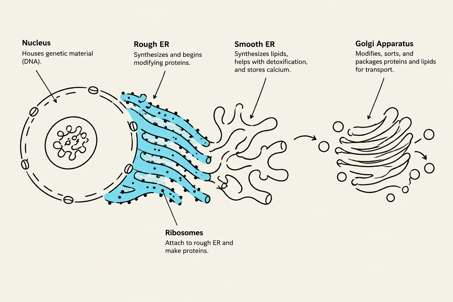
The ER is the largest membrane-bound organelle and is physically continuous with the nuclear envelope. It is divided into two distinct functional regions:
Rough ER: Studded with ribosomes, this region is the primary site for the synthesis of proteins destined for secretion, the plasma membrane, or for use within other organelles like lysosomes. It also performs glycosylation, the process of adding sugar chains to proteins.
Smooth ER: Lacking ribosomes, the smooth ER specializes in lipid synthesis (such as phospholipids and steroids), carbohydrate metabolism, and detoxification. In muscle cells, a specialized form called the sarcoplasmic reticulum serves as a vital reservoir for calcium ions.
To ensure cellular health, the ER employs "quality control" mechanisms. Chaperone proteins assist in proper folding, while the Unfolded Protein Response (UPR) can halt protein translation if misfolded proteins accumulate to dangerous levels.
Diagram illustrating the nucleus, rough endoplasmic reticulum (ER), smooth ER, ribosomes, and Golgi apparatus, key organelles within a eukaryotic cell involved in protein and lipid synthesis, modification, and transport.
The Golgi Apparatus and Lysosomes
Once proteins and lipids leave the ER via transitional vesicles, they arrive at the Golgi apparatus. The Golgi acts as the cell's "post office," further modifying proteins through glycosylation and sorting them into specific vesicles for delivery to their final destinations.
For waste management, the cell utilizes lysosomes. These organelles contain acid hydrolases—enzymes that function optimally in acidic environments—to degrade cellular waste, including old organelles through a process called autophagy.
Peroxisomes
While often grouped with the endomembrane system, peroxisomes have a specialized role in oxidative metabolism. They break down long-chain fatty acids and perform detoxification tasks. A byproduct of these reactions is hydrogen peroxide (H2O2), which peroxisomes safely decompose using the enzyme catalase.
Exam Tip: Remember the distinction between the ER types. If a question mentions "steroid synthesis" or "detoxification," think Smooth ER. If it mentions "secretory proteins" or "ribosomes," think Rough ER.
Microscopy: Visualizing the Subcellular World
To study these organelles, biologists rely on the physics of light and electrons. The quality of any microscopic image is determined by two primary factors: magnification and resolution.
Resolution and Numerical Aperture
While magnification makes an object appear larger, resolution (D) is the most critical property of a microscope. Resolution is the ability to distinguish two closely positioned objects as separate entities. A smaller value for D indicates better resolution.
The resolution of a light microscope is mathematically constrained by the wavelength of light (λ) and the Numerical Aperture (NA) of the lens. The relationship is expressed as:
D=nsinα0.61λ
Where:
λ is the wavelength of light.
n is the refractive index of the medium (e.g., air or oil).
α is the half-angle of the cone of light entering the objective lens.
Increasing the Numerical Aperture (by using a higher α or adding immersion oil to increase n) decreases D, thereby improving the resolution.
Microscopy Techniques
Different techniques are chosen based on the specimen's nature:
Brightfield Microscopy: The simplest form, where the specimen is viewed against a bright background.
Phase-Contrast Microscopy: Ideal for observing live, transparent cells by utilizing differences in refractive indices to create contrast.
Fluorescence Microscopy: Uses fluorescent dyes that absorb light at an excitation wavelength and emit it at a longer wavelength, allowing specific molecules to be tagged and visualized against a dark background.
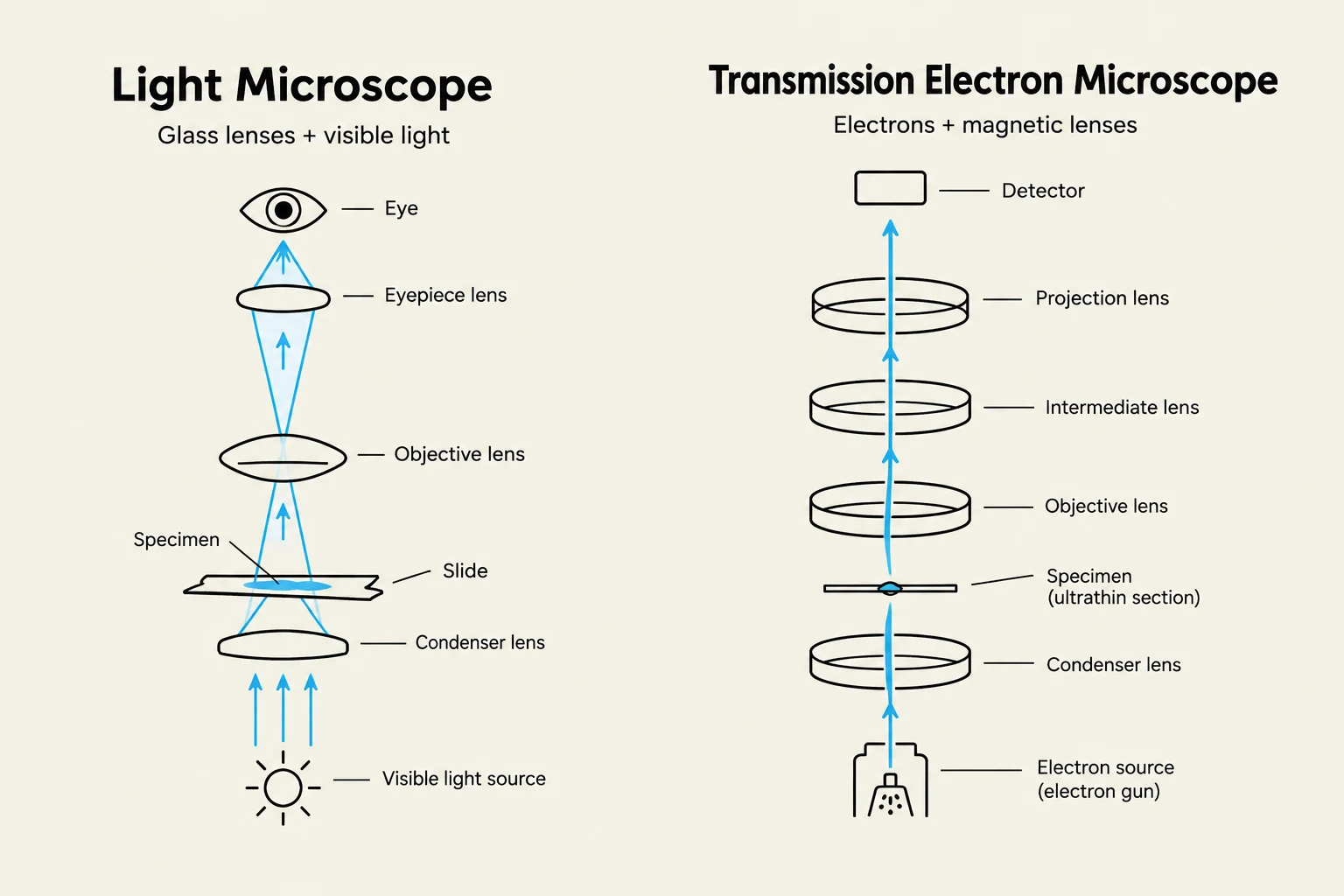
Transmission Electron Microscopy (TEM): Instead of light, TEM uses a beam of electrons. Because the wavelength of an electron is significantly shorter than that of visible light, TEM can achieve a resolution up to 100,000 times greater than a light microscope, reaching scales as small as 0.2 nm.
Diagrams comparing the components and operation of a light microscope and a transmission electron microscope.
Subcellular Fractionation: Isolating the Components
When researchers want to study the specific enzymes or proteins within an organelle, they must first isolate that organelle from the rest of the cell through subcellular fractionation.
The Fractionation Process
The process begins with homogenization, where cells are disrupted (using a blender, sonication, or mechanical grinding) in an isotonic solution to create a "homogenate". This must be done on ice to prevent the degradation of proteins and organelles.
Differential Centrifugation: This method separates organelles based on their size and density using increasing speeds of centrifugal force.
Low speed (1,000 x g) pellets the large, dense nuclei.
Medium speed (20,000 x g) pellets mitochondria, peroxisomes, and lysosomes.
High speed (100,000 x g) pellets microsomes (ER fragments) and plasma membrane vesicles.
Equilibrium Density-Gradient Centrifugation: To achieve higher purity, organelles are layered onto a sucrose gradient. During centrifugation, each organelle migrates until it reaches a position where its density matches the density of the surrounding sucrose solution.
Biochemical Verification
Once an organelle is isolated, its purity must be verified. Researchers often use Western blot analysis to detect the presence of specific "marker proteins" that are known to reside only in the target organelle. For example, if a researcher is studying lysosomes, they would use antibodies to check for the presence of lysosomal acid hydrolases in their sample.
Diagram illustrating the process of differential centrifugation to separate cellular components like nuclei, mitochondria, microsomes, and cytosol based on their sedimentation rates at different centrifugal forces and times.
Summary Table for Centrifugation
Step
Force (x g)
Primary Result (Pellet)
1
1,000
Nuclei
2
20,000
Mitochondria, Lysosomes, Peroxisomes
3
100,000
Microsomes (ER), Plasma Membrane
Keep reading for free.
Create an account to finish this reading and make it part of your study routine.