Cellular Interactions and Transport Across the Plasma Membrane
Explores the extracellular environment, including body fluids and the extracellular matrix, and details various transport mechanisms across the plasma membrane, such as passive and active transport, and their physiological significance.
Cells do not exist in isolation; they are constantly interacting with their surroundings to obtain nutrients, expel waste, and communicate with one another. This surrounding area, known as the extracellular environment, is a complex medium that provides both the chemical signals and the physical structure necessary for life to thrive.
The Extracellular Environment
The extracellular environment is composed primarily of water. It can be divided into two main components: body fluids and the extracellular matrix.
Body Fluids
Of the total water in the human body, approximately 33% is extracellular. This is further subdivided into:
Blood Plasma: Makes up about 20% of the extracellular fluid (which is 8% of total body water).
Interstitial Fluid: The fluid located between the cells of tissues, acting as a bridge to the blood plasma. It accounts for 80% of the extracellular fluid (25% of total body water).
The Extracellular Matrix (ECM)
While fluids provide the medium for transport, the extracellular matrix provides the structural scaffolding. The ECM consists of a gel-like ground substance made of glycoproteins and proteoglycans, reinforced by protein fibers such as collagen and elastin. Cells attach to this matrix using specialized glycoproteins called integrins, which extend from the cell's cytoskeleton to facilitate adhesion, motility, and proliferation.
The Plasma Membrane and Transport Mechanisms
The gateway between the cell and its environment is the plasma membrane. This membrane is selectively permeable, meaning it regulates what enters and exits the cell. Generally, the membrane restricts large molecules like proteins and nucleic acids, while allowing the passage of ions, nutrients (such as glucose and amino acids), and wastes.
Transport across this membrane is categorized by whether or not it requires metabolic energy (ATP).
Passive Transport
In passive transport, molecules move "down" their concentration gradient—from an area of high concentration to an area of low concentration—without the expenditure of energy.
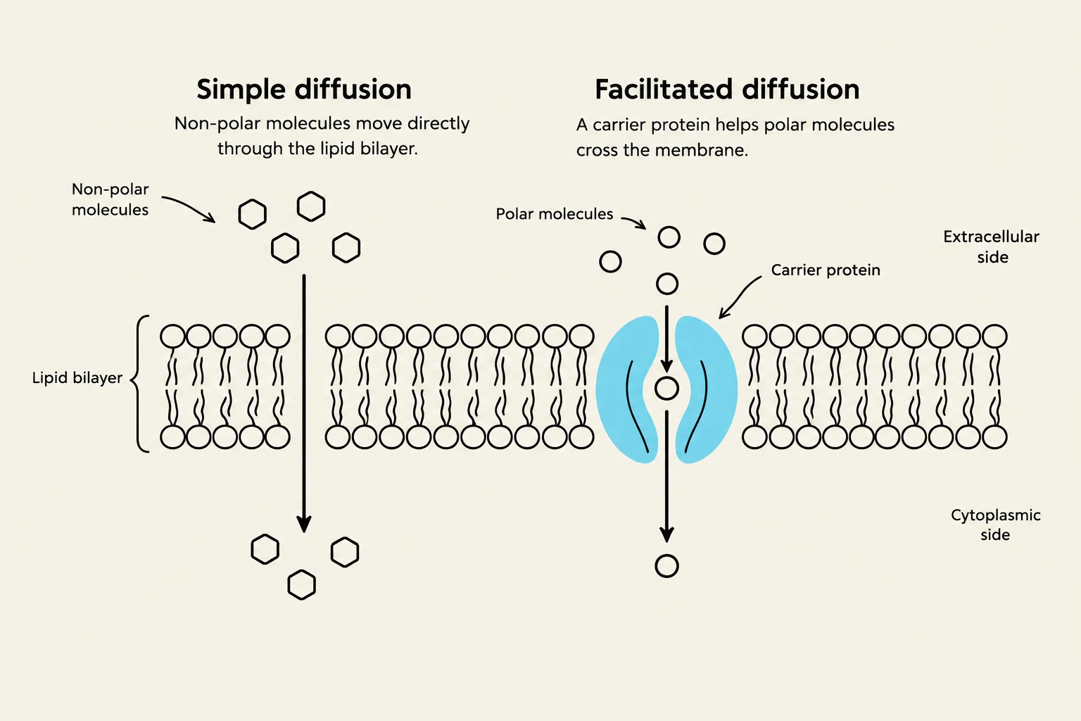
Simple Diffusion: Non-polar, lipid-soluble molecules (like oxygen, carbon dioxide, and steroid hormones) can pass directly through the phospholipid bilayer. Small ions and polar molecules can also move through the membrane via protein channels.
Facilitated Diffusion: Large or polar organic molecules, such as glucose or amino acids, cannot cross the lipid bilayer alone. They require specific carrier proteins that undergo a conformational change to move the molecule across.
Osmosis: This is the unique diffusion of the solvent (water). Water moves from an area of low solute concentration to an area of high solute concentration. While water can move slowly directly through the membrane, it moves much faster through specialized protein channels called aquaporins.
Exam Tip: The rate of diffusion is not constant. It increases with a larger concentration difference, higher temperatures, and a larger membrane surface area (such as that provided by microvilli).
This diagram illustrates two types of diffusion across a cell membrane: simple diffusion for non-polar molecules and facilitated diffusion for polar molecules using a carrier protein.
Active Transport
Active transport moves substances "uphill," against their concentration gradient (from low to high concentration). This process requires energy in the form of ATP and specific carrier proteins known as pumps.
Primary Active Transport: The hydrolysis of ATP directly powers the pump. A classic example is the Na+/K+ pump, which pumps three Na+ ions out of the cell and two K+ ions into the cell. This helps maintain osmotic balance and electrical gradients. Another example is the Ca2+ pump, which maintains low cytoplasmic calcium levels.
Secondary Active Transport: This is also called coupled transport. It uses the energy stored in the gradient of one molecule (usually Na+ moving down its gradient) to drive another molecule "uphill". This can occur via symport (both molecules move in the same direction, like glucose and sodium) or antiport (molecules move in opposite directions).
Quantifying Solutions: Molarity, Molality, and Osmolality
To understand how solutes affect cells, scientists use different ways to measure concentration.
Molarity (M): The number of moles of solute per liter of solution.
Molality (m): The ratio of moles of solute to kilograms of water.
Osmolality: The total concentration of all osmotically active particles in a solution.
In medicine, osmolality is often preferred because it is less affected by changes in temperature and pressure. It is crucial to remember that electrolytes dissociate in water. For example, a 1.0 m solution of NaCl will have an osmolality of 2 Osm because it dissociates into two particles: Na+ and Cl−.
Osmotic Pressure and Tonicity
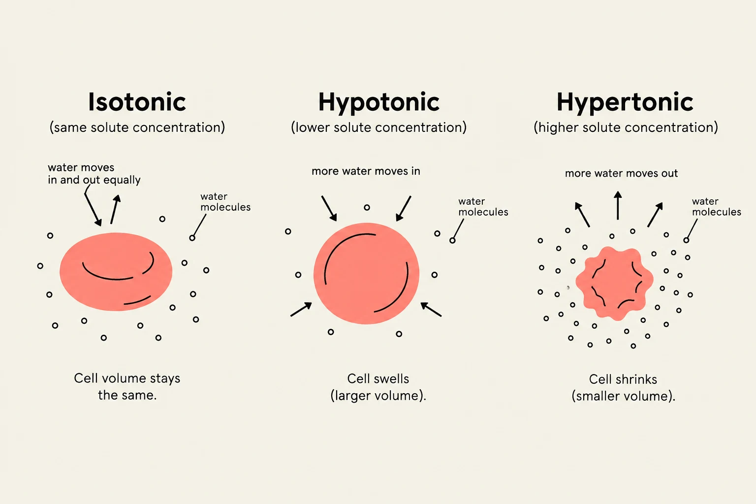
Osmotic pressure is the force required to stop the movement of water via osmosis. While osmolality describes the concentration of a solution, tonicity describes how that solution affects cell volume by driving water movement.
Isotonic: The solute concentration is the same as the cell; no net water movement occurs.
Hypotonic: The solution has a lower solute concentration than the cell; water moves into the cell, causing it to swell.
Hypertonic: The solution has a higher solute concentration than the cell; water moves out of the cell, causing it to shrink.
Diagram illustrating the effects of isotonic, hypotonic, and hypertonic solutions on a cell, showing water movement and resulting cell volume changes.
Homeostasis and Physiological Regulation
The body must maintain a constant plasma osmolality to prevent damage to sensitive cells, such as neurons. This is achieved through a homeostatic feedback loop involving the hypothalamus:
Detection: Osmoreceptors in the hypothalamus detect an increase in blood osmolality (indicating dehydration).
Response: The brain triggers the sensation of thirst and signals the release of antidiuretic hormone (ADH).
Action: ADH causes the insertion of more aquaporins into the kidney tubule membranes, increasing water reabsorption back into the blood and decreasing urine output.
When osmolality returns to normal, ADH release decreases, and water excretion increases, completing the cycle.
Summary Table: Transport Comparison
Feature
Simple Diffusion
Facilitated Diffusion
Primary Active Transport
Secondary Active Transport
Gradient Direction
High → Low
High → Low
Low → High
Low → High
Energy (ATP) Required?
No
No
Yes (Directly)
Yes (Indirectly)
Requires Protein?
No
Yes (Carrier/Channel)
Yes (Pump)
Yes (Cotransporter)
Example
O2,CO2
Glucose (via GLUT)
Na+/K+ Pump
Glucose/Na+ Symport
Keep reading for free.
Create an account to finish this reading and make it part of your study routine.