Cellular Injury, Adaptation, and Death: Mechanisms and Pathways
An in-depth explanation of how cells respond to stress, including adaptation, reversible and irreversible injury, and the distinct processes of necrosis and apoptosis. Covers clinical contexts like hypoxia and ischemia.
Cells are dynamic units that constantly strive to maintain a stable internal environment known as homeostasis. When cells encounter physiological stress or pathological stimuli, they do not immediately die. Instead, they undergo a series of responses designed to preserve viability. This article explores the spectrum of cellular responses, ranging from beneficial adaptations to the catastrophic processes of cell death.
Cellular Adaptation: The First Line of Defense
When a cell encounters a stressor—such as increased workload, decreased nutrition, or changes in hormone levels—it often attempts to adapt. Adaptation involves reversible changes in the size, number, phenotype, metabolic activity, or functions of cells. If the stress is removed, the cell can often return to its original state; however, if the stress exceeds the cell's adaptive capacity, cellular injury occurs.
Common forms of cellular adaptation include:
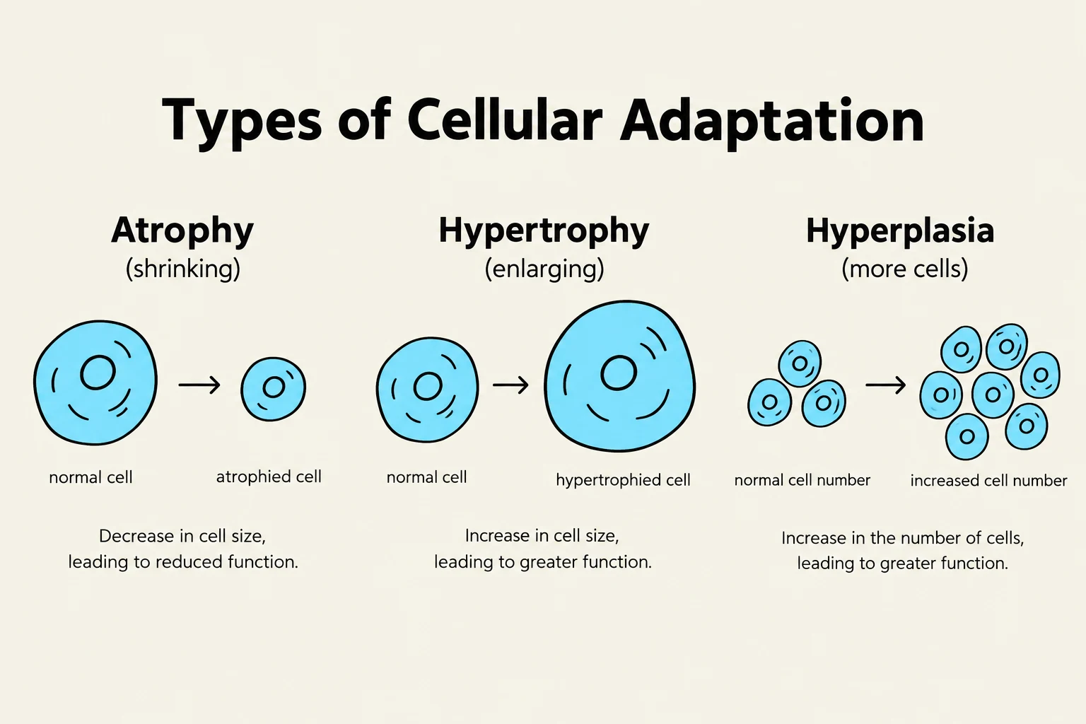
Atrophy: A decrease in cell size and number, often resulting in a reduction in the size of an organ. This can be physiological (e.g., the shrinking of the uterus after childbirth) or pathological (e.g., muscle wasting due to disuse).
Hypertrophy: An increase in the size of cells, leading to an increase in the size of the organ. This is common in cells that cannot divide, such as cardiac muscle cells responding to hypertension.
Hyperplasia: An increase in the number of cells in an organ or tissue. This can be physiological (e.g., hormonal stimulation of breast tissue) or pathological (e.g., endometrial hyperplasia).
Metaplasia: A reversible change in which one adult cell type is replaced by another adult cell type. A classic example is the change of ciliated columnar epithelium to squamous epithelium in the airways of chronic smokers.
Dysplasia: Characterized by disordered growth and loss of cellular uniformity. While not a true adaptation, it is often a precursor to neoplasia (cancer).
Exam Tip: Distinguish between hypertrophy and hyperplasia. Hypertrophy is an increase in size (common in permanent cells like muscle), whereas hyperplasia is an increase in number (common in cells capable of division).
This diagram illustrates three types of cellular adaptation: atrophy (shrinking), hypertrophy (enlarging), and hyperplasia (more cells), showing the changes in cell size or number and their functional consequences.
Cellular Injury: Reversible vs. Irreversible
When the stress is too severe or persistent, the cell can no longer maintain homeostasis, leading to cellular injury. This injury is categorized into two distinct stages: reversible and irreversible.
Reversible Injury
Reversible injury represents a stage where the cell can return to its normal state if the injurious stimulus is withdrawn. The primary hallmarks of reversible injury include:
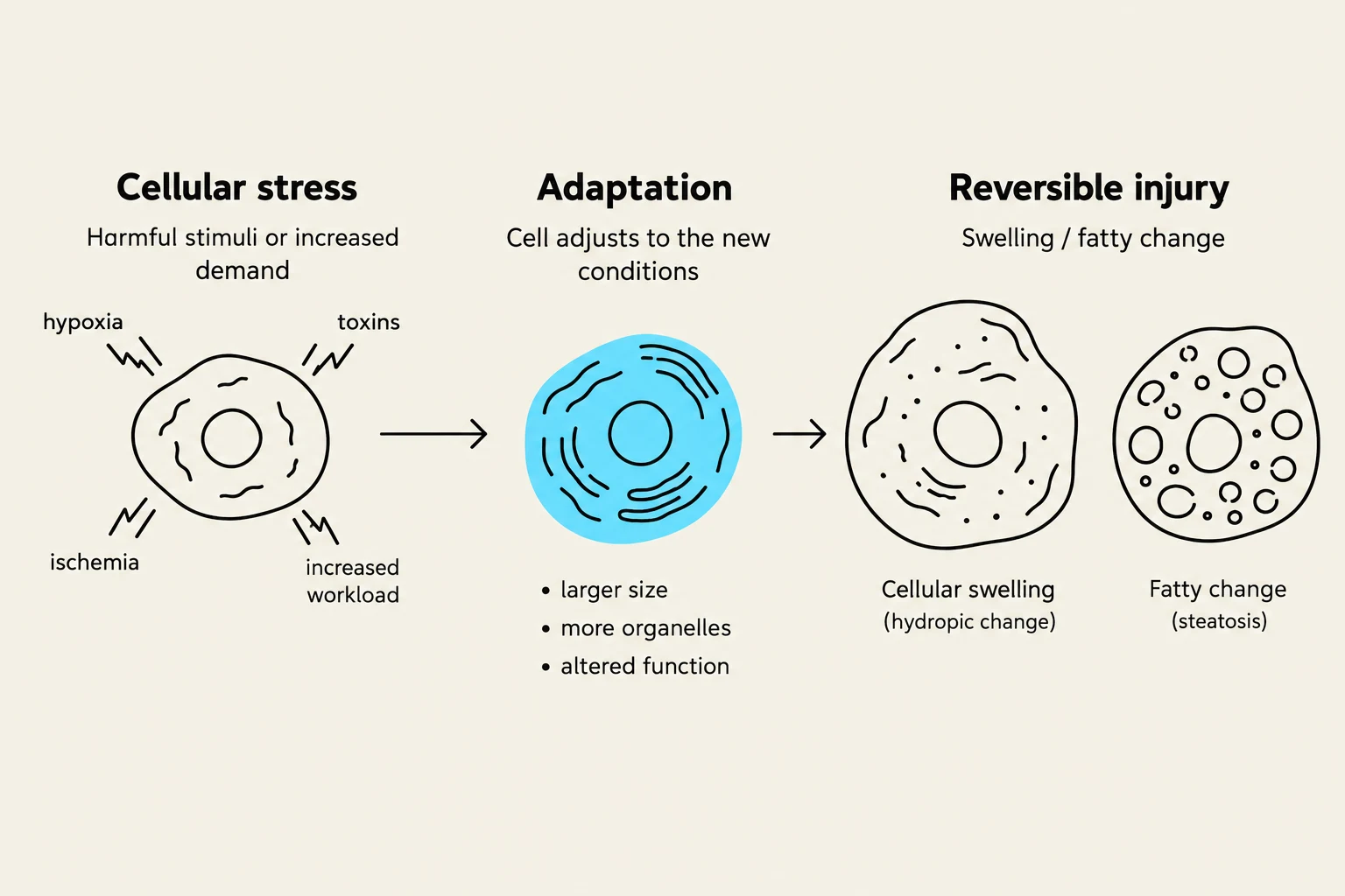
Cellular Swelling (Hydropic Change): This occurs when the cell's energy-dependent ion pumps (such as the Na+/K+ ATPase pump) fail due to ATP depletion. This failure leads to an influx of sodium and water into the cell, causing it to swell.
Fatty Change (Steatosis): This is the appearance of lipid vacuoles within the cytoplasm, frequently observed in organs like the liver when metabolic pathways are disrupted.
Irreversible Injury
If the stimulus persists, the cell reaches a "point of no return," transitioning into irreversible injury and eventual cell death. The critical mechanisms driving this transition include:
Mitochondrial Dysfunction: Permanent damage to the mitochondria prevents the cell from generating sufficient ATP to sustain life.
Membrane Damage: Loss of integrity in the plasma membrane, lysosomal membranes (releasing digestive enzymes), and mitochondrial membranes.
Calcium Influx: A massive, uncontrolled increase in intracellular calcium levels activates various enzymes (proteases, nucleases, and phospholipases) that actively degrade cellular components.
This diagram illustrates the process of cellular stress leading to adaptation or reversible injury, such as cellular swelling and fatty change.
Mechanisms of Cell Death
Cell death is the final outcome of irreversible injury. Depending on the mechanism and the context, cell death follows one of two primary pathways: necrosis or apoptosis.
Necrosis: The Pathological Process
Necrosis is always a pathological process characterized by cell swelling, protein denaturation, and the breakdown of organelles. The defining feature of necrosis is the rupture of the plasma membrane. When the membrane breaks, intracellular contents leak into the surrounding tissue, which triggers an inflammatory response.
Common forms of necrosis include:
Coagulative necrosis: Often seen in infarcts (ischemic death) in most organs except the brain.
Liquefactive necrosis: Typical of focal bacterial or fungal infections and brain infarcts.
Caseous necrosis: Characterized by a "cheese-like" appearance, often seen in tuberculosis.
Apoptosis: Programmed Cell Death
Unlike necrosis, apoptosis is a highly regulated, energy-dependent form of "programmed cell death". It can be physiological (e.g., removing cells during embryonic development or eliminating self-reactive immune cells) or pathological (e.g., following DNA damage).
In apoptosis, the cell shrinks and fragments into small, membrane-bound vesicles called apoptotic bodies. These bodies are efficiently phagocytosed by neighboring cells or macrophages without releasing their contents into the extracellular space, thereby avoiding an inflammatory response.
This diagram illustrates the distinct processes of necrosis and apoptosis, two forms of cell death. Necrosis involves cell swelling, membrane rupture, and inflammation, leading to the release of cell contents and the arrival of immune cells. Apoptosis, in contrast, is a controlled process characterized by cell shrinkage and fragmentation into apoptotic bodies without inflammation.
Clinical Context: Hypoxia and Ischemia
To understand these processes in a clinical setting, we must distinguish between two common causes of cellular injury: hypoxia and ischemia.
Hypoxia refers to a deficiency in oxygen supply to the tissues. This can be caused by various factors, such as anemia or respiratory failure.
Ischemia is a more severe form of injury caused by a reduction in blood supply to a tissue. Because blood carries not only oxygen but also nutrients (like glucose) and removes metabolic waste, ischemia is generally more damaging than hypoxia alone.
For example, in a myocardial infarction (heart attack), the blockage of a coronary artery causes ischemia, which rapidly leads to hypoxia, then reversible injury, and finally necrosis of the cardiac muscle if blood flow is not restored.
Summary Table: Necrosis vs. Apoptosis
Feature
Necrosis
Apoptosis
Cell Size
Enlarged (swelling)
Reduced (shrinkage)
Nucleus
Pyknosis → Karyorrhexis → Karyolysis
Fragmentation
Plasma Membrane
Ruptured
Intact (blebbing)
Inflammation
Frequent/Significant
None
Physiological?
Always Pathological
Can be Physiological
Keep reading for free.
Create an account to finish this reading and make it part of your study routine.